5LTQ image
Deposition Date 2016-09-07
Release Date 2016-12-07
Last Version Date 2024-11-13
Entry Detail
PDB ID:
5LTQ
Title:
Structure of the Yellow Fluorescent Protein lanYFP from Branchiostoma lanceolatum at pH 7.5
Biological Source:
Source Organism:
Method Details:
Experimental Method:
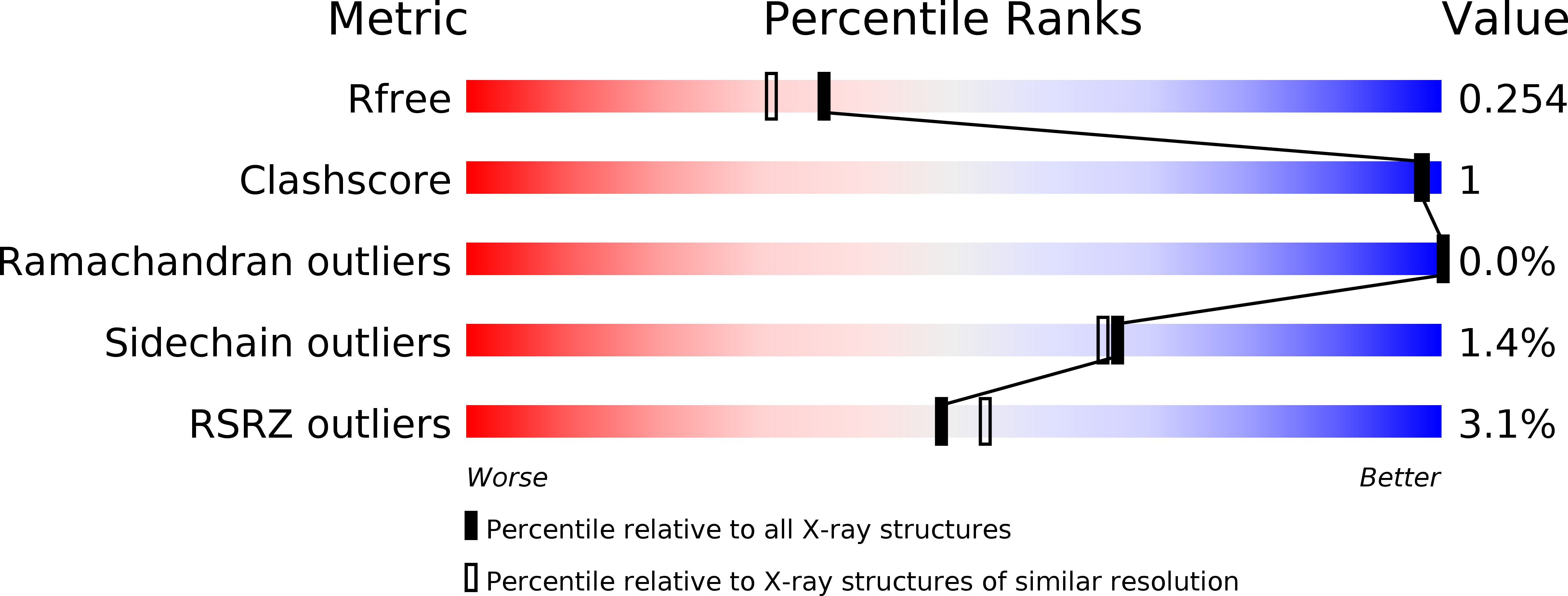
Resolution:
2.05 Å
R-Value Free:
0.25
R-Value Work:
0.22
R-Value Observed:
0.22
Space Group:
P 1 21 1
Macromolecular Entities
Structures with similar UniProt ID
Protein Blast
Polymer Type:polypeptide(L)
Molecule:Green fluorescent protein blFP-Y3
Gene (Uniprot):blFP-Y3
Chain IDs:A, B (auth: D), C (auth: B), D (auth: C), E, F (auth: H), G (auth: F), H (auth: G), I, J (auth: L), K (auth: J), L (auth: K), M, N (auth: P), O (auth: N), P (auth: O)
Chain Length:269
Number of Molecules:16
Biological Source:Branchiostoma lanceolatum
Modified Residue
Compound ID Chain ID Parent Comp ID Details 2D Image
CR2 A GLY chromophore
Ligand Molecules
Primary Citation
Structural analysis of the bright monomeric yellow-green fluorescent protein mNeonGreen obtained by directed evolution.
Acta Crystallogr D Struct Biol 72 1298 1307 (2016)
PMID: 27917830 DOI: 10.1107/S2059798316018623

Abstact

Until recently, genes coding for homologues of the autofluorescent protein GFP had only been identified in marine organisms from the phyla Cnidaria and Arthropoda. New fluorescent-protein genes have now been found in the phylum Chordata, coding for particularly bright oligomeric fluorescent proteins such as the tetrameric yellow fluorescent protein lanYFP from Branchiostoma lanceolatum. A successful monomerization attempt led to the development of the bright yellow-green fluorescent protein mNeonGreen. The structures of lanYFP and mNeonGreen have been determined and compared in order to rationalize the directed evolution process leading from a bright, tetrameric to a still bright, monomeric fluorescent protein. An unusual discolouration of crystals of mNeonGreen was observed after X-ray data collection, which was investigated using a combination of X-ray crystallography and UV-visible absorption and Raman spectroscopies, revealing the effects of specific radiation damage in the chromophore cavity. It is shown that X-rays rapidly lead to the protonation of the phenolate O atom of the chromophore and to the loss of its planarity at the methylene bridge.

Legend

Protein

Chemical

Disease

Primary Citation of related structures