Deposition Date
2016-09-05
Release Date
2017-01-18
Last Version Date
2024-01-17
Entry Detail
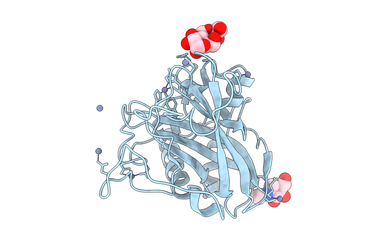
Biological Source:
Source Organism(s):
Aspergillus oryzae RIB40 (Taxon ID: 510516)
Expression System(s):
Method Details:
Experimental Method:
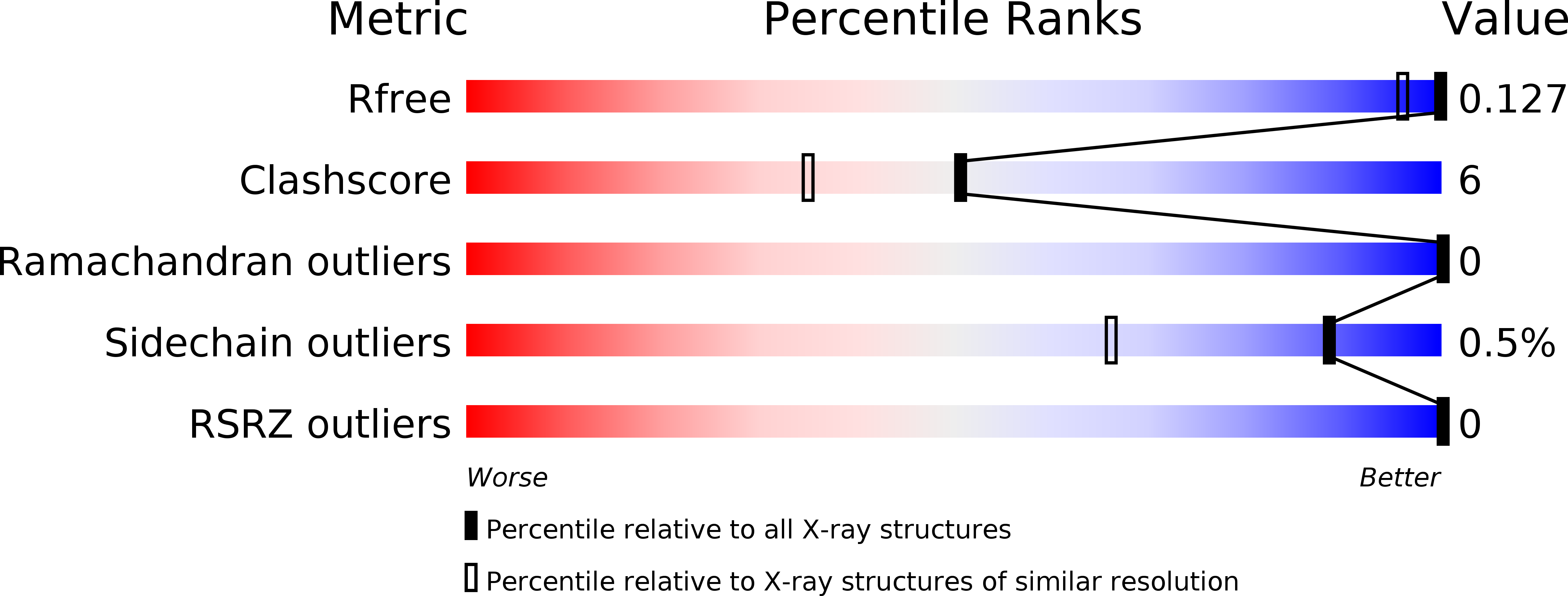
Resolution:
1.10 Å
R-Value Free:
0.12
R-Value Work:
0.10
R-Value Observed:
0.10
Space Group:
P 21 21 21