5LNQ image
Entry Detail
PDB ID:
5LNQ
Keywords:
Title:
Crystal structure of SCP2 thiolase from Leishmania mexicana. Complex of the C123A mutant with acetoacetyl-CoA.
Biological Source:
Source Organism:
PDB Version:
Deposition Date:
2016-08-06
Release Date:
2017-01-18
Method Details:
Experimental Method:
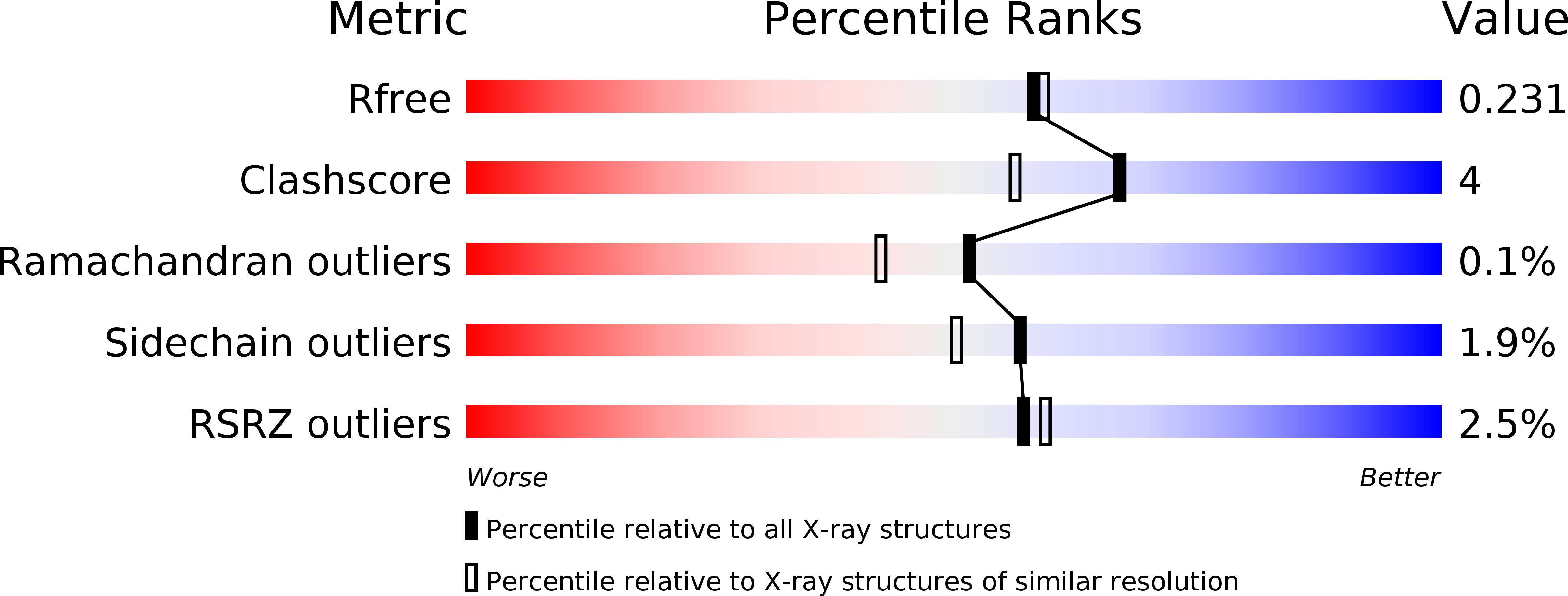
Resolution:
1.98 Å
R-Value Free:
0.22
R-Value Work:
0.18
R-Value Observed:
0.19
Space Group:
P 1 21 1
Macromolecular Entities
Polymer Type:polypeptide(L)
Description:3-ketoacyl-CoA thiolase-like protein
Mutations:C123A
Chain IDs:A, B, C, D
Chain Length:457
Number of Molecules:4
Biological Source:Leishmania mexicana
Ligand Molecules
Primary Citation
Crystallographic substrate binding studies of Leishmania mexicana SCP2-thiolase (type-2): unique features of oxyanion hole-1.
Protein Eng. Des. Sel. 30 225 233 (2017)
PMID: 28062645 DOI: 10.1093/protein/gzw080

Abstact

Structures of the C123A variant of the dimeric Leishmania mexicana SCP2-thiolase (type-2) (Lm-thiolase), complexed with acetyl-CoA and acetoacetyl-CoA, respectively, are reported. The catalytic site of thiolase contains two oxyanion holes, OAH1 and OAH2, which are important for catalysis. The two structures reveal for the first time the hydrogen bond interactions of the CoA-thioester oxygen atom of the substrate with the hydrogen bond donors of OAH1 of a CHH-thiolase. The amino acid sequence fingerprints (xS, EAF, G P) of three catalytic loops identify the active site geometry of the well-studied CNH-thiolases, whereas SCP2-thiolases (type-1, type-2) are classified as CHH-thiolases, having as corresponding fingerprints xS, DCF and G P. In all thiolases, OAH2 is formed by the main chain NH groups of two catalytic loops. In the well-studied CNH-thiolases, OAH1 is formed by a water (of the Wat-Asn(NEAF) dyad) and NE2 (of the GHP-histidine). In the two described liganded Lm-thiolase structures, it is seen that in this CHH-thiolase, OAH1 is formed by NE2 of His338 (HDCF) and His388 (GHP). Analysis of the OAH1 hydrogen bond networks suggests that the GHP-histidine is doubly protonated and positively charged in these complexes, whereas the HDCF histidine is neutral and singly protonated.

Legend

Protein

Chemical

Disease

Primary Citation of related structures