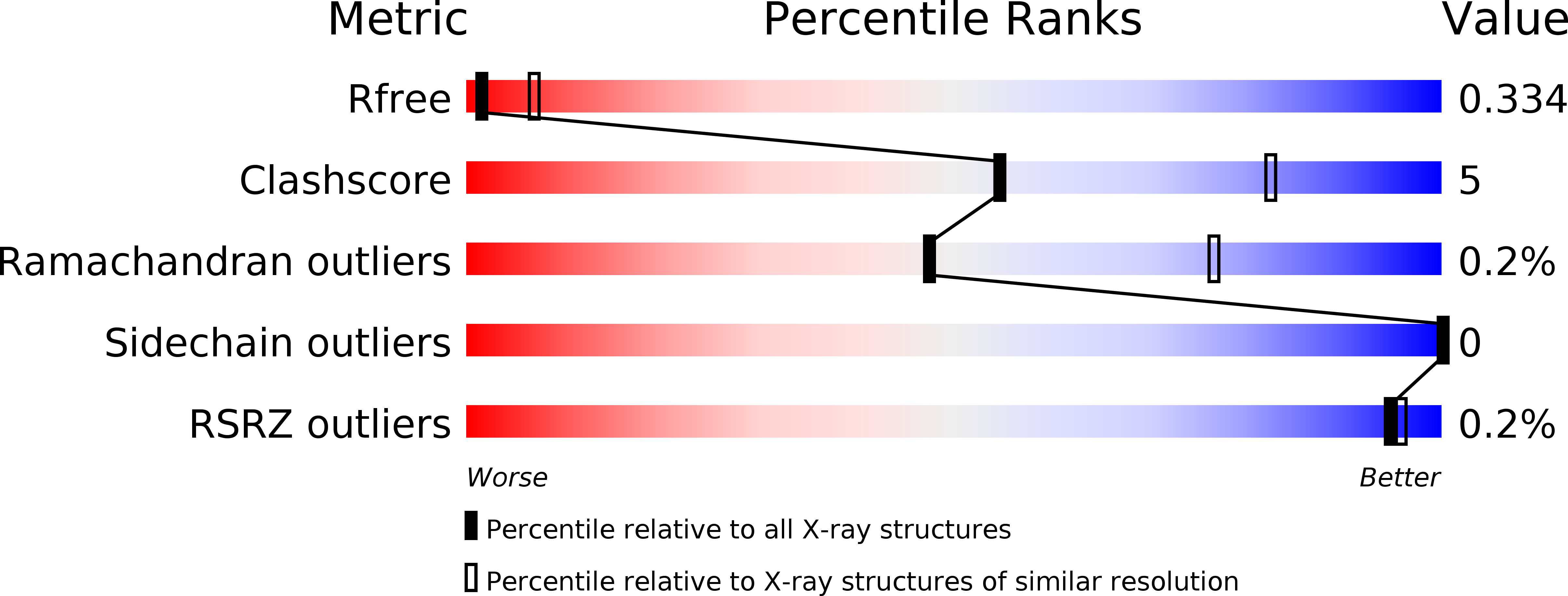
5LNL image
Deposition Date 2016-08-05
Release Date 2017-02-15
Last Version Date 2024-01-10
Entry Detail
PDB ID:
5LNL
Keywords:
Title:
Crystal structure of Hsf 1608-1749 putative domain 1
Biological Source:
Source Organism:
Method Details:
Experimental Method:
Resolution:
3.30 Å
R-Value Free:
0.33
R-Value Work:
0.29
Space Group:
C 1 2 1
Macromolecular Entities
Structures with similar UniProt ID
Protein Blast
Polymer Type:polypeptide(L)
Molecule:Hsf
Gene (Uniprot):hsf
Chain IDs:A, B, C, D, E, F, G, H, I
Chain Length:151
Number of Molecules:9
Biological Source:Haemophilus influenzae
Ligand Molecules
Primary Citation
The crystal structure of PD1, a Haemophilus surface fibril domain.
Acta Crystallogr F Struct Biol Commun 73 101 108 (2017)
PMID: 28177321 DOI: 10.1107/S2053230X17001406

Abstact

The Haemophilus surface fibril (Hsf) is an unusually large trimeric autotransporter adhesin (TAA) expressed by the most virulent strains of H. influenzae. Hsf is known to mediate adhesion between pathogen and host, allowing the establishment of potentially deadly diseases such as epiglottitis, meningitis and pneumonia. While recent research has suggested that this TAA might adopt a novel `hairpin-like' architecture, the characterization of Hsf has been limited to in silico modelling and electron micrographs, with no high-resolution structural data available. Here, the crystal structure of Hsf putative domain 1 (PD1) is reported at 3.3 Å resolution. The structure corrects the previous domain annotation by revealing the presence of an unexpected N-terminal TrpRing domain. PD1 represents the first Hsf domain to be solved, and thus paves the way for further research on the `hairpin-like' hypothesis.

Legend

Protein

Chemical

Disease

Primary Citation of related structures