5LGX image
Deposition Date 2016-07-08
Release Date 2017-02-15
Last Version Date 2024-01-10
Entry Detail
PDB ID:
5LGX
Keywords:
Title:
Structure of Oxidised Pentaerythritol Tetranitrate Reductase
Biological Source:
Source Organism:
Host Organism:
Method Details:
Experimental Method:
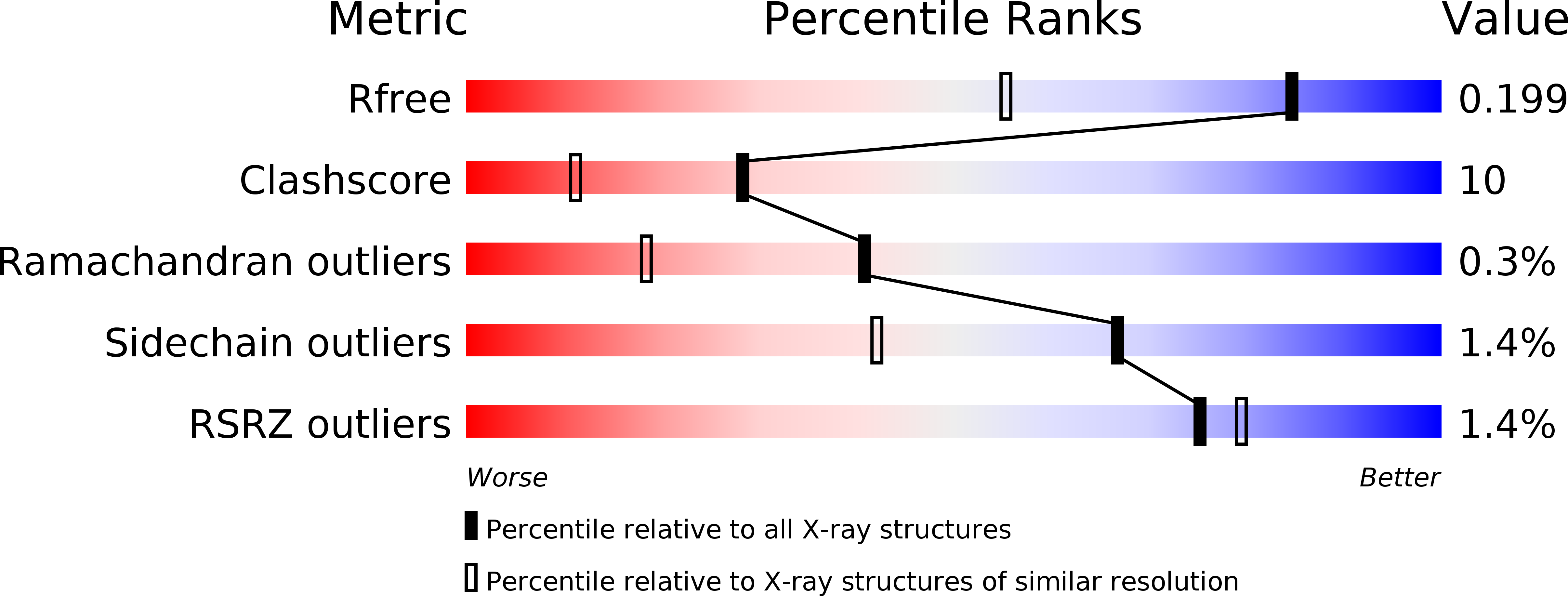
Resolution:
1.50 Å
R-Value Free:
0.20
R-Value Work:
0.15
R-Value Observed:
0.15
Space Group:
P 21 21 21
Macromolecular Entities
Polymer Type:polypeptide(L)
Molecule:Pentaerythritol tetranitrate reductase
Gene (Uniprot):onr
Chain IDs:A
Chain Length:363
Number of Molecules:1
Biological Source:Enterobacter cloacae
Primary Citation
Combining X-ray and neutron crystallography with spectroscopy.
Acta Crystallogr D Struct Biol 73 141 147 (2017)
PMID: 28177310 DOI: 10.1107/S2059798316016314

Abstact

X-ray protein crystallography has, through the determination of the three-dimensional structures of enzymes and their complexes, been essential to the understanding of biological chemistry. However, as X-rays are scattered by electrons, the technique has difficulty locating the presence and position of H atoms (and cannot locate H+ ions), knowledge of which is often crucially important for the understanding of enzyme mechanism. Furthermore, X-ray irradiation, through photoelectronic effects, will perturb the redox state in the crystal. By using single-crystal spectrophotometry, reactions taking place in the crystal can be monitored, either to trap intermediates or follow photoreduction during X-ray data collection. By using neutron crystallography, the positions of H atoms can be located, as it is the nuclei rather than the electrons that scatter neutrons, and the scattering length is not determined by the atomic number. Combining the two techniques allows much greater insight into both reaction mechanism and X-ray-induced photoreduction.

Legend

Protein

Chemical

Disease

Primary Citation of related structures