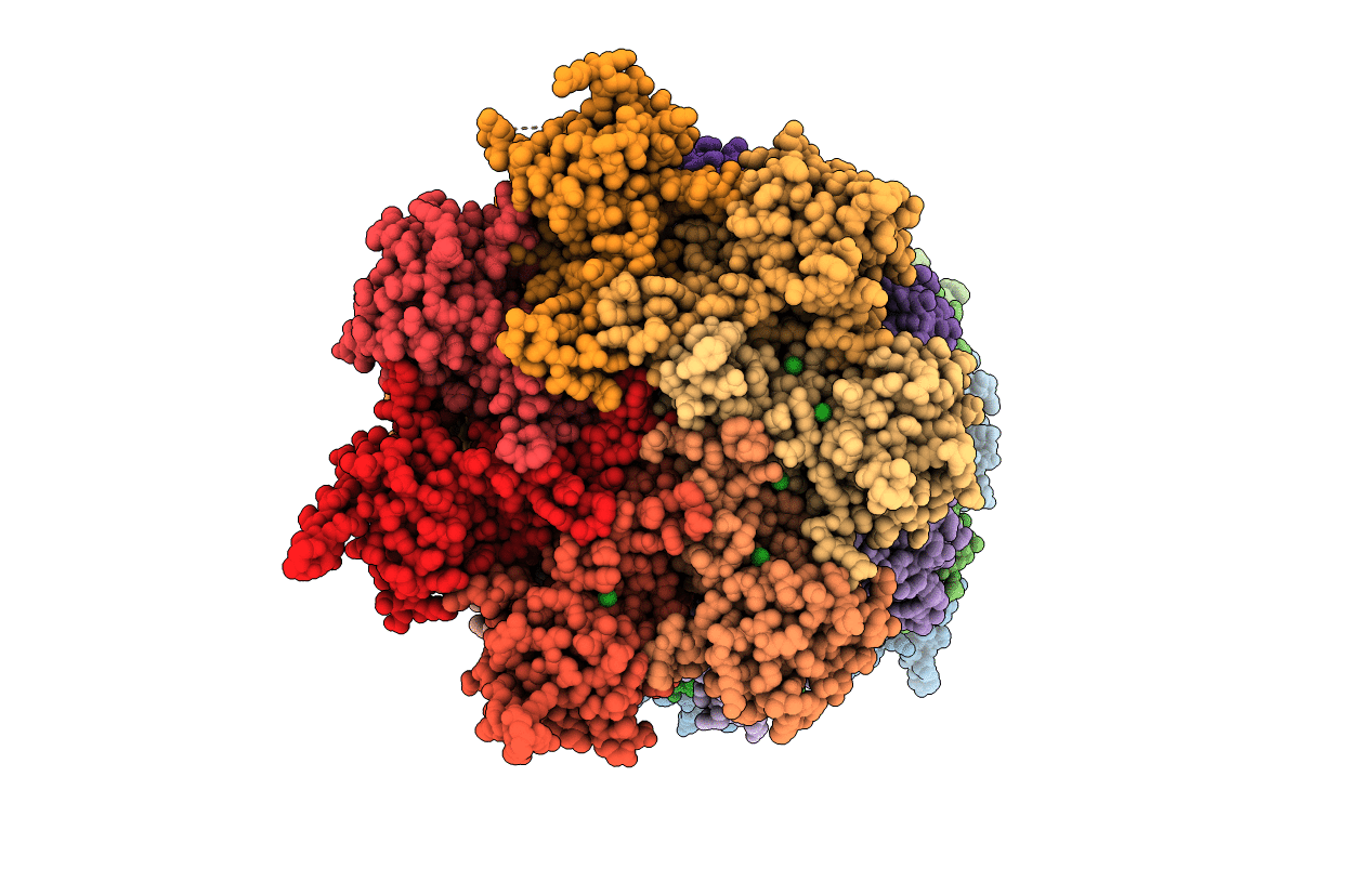
5LF7 image
Deposition Date 2016-06-30
Release Date 2016-08-17
Last Version Date 2024-01-10
Entry Detail
PDB ID:
5LF7
Keywords:
Title:
Human 20S proteasome complex with Ixazomib at 2.0 Angstrom
Biological Source:
Source Organism:
Homo sapiens (Taxon ID: 9606)
Method Details:
Experimental Method:
Resolution:
2.00 Å
R-Value Free:
0.21
R-Value Work:
0.17
R-Value Observed:
0.17
Space Group:
P 21 21 21
Macromolecular Entities
Polymer Type:polypeptide(L)
Molecule:Proteasome subunit alpha type-2
Gene (Uniprot):PSMA2
Chain IDs:A, O
Chain Length:234
Number of Molecules:2
Biological Source:Homo sapiens
Polymer Type:polypeptide(L)
Molecule:Proteasome subunit alpha type-4
Gene (Uniprot):PSMA4
Chain IDs:B, P
Chain Length:261
Number of Molecules:2
Biological Source:Homo sapiens
Polymer Type:polypeptide(L)
Molecule:Proteasome subunit alpha type-7
Gene (Uniprot):PSMA7
Chain IDs:C, Q
Chain Length:248
Number of Molecules:2
Biological Source:Homo sapiens
Polymer Type:polypeptide(L)
Molecule:Proteasome subunit alpha type-5
Gene (Uniprot):PSMA5
Chain IDs:D, R
Chain Length:241
Number of Molecules:2
Biological Source:Homo sapiens
Polymer Type:polypeptide(L)
Molecule:Proteasome subunit alpha type-1
Gene (Uniprot):PSMA1
Chain IDs:E, S
Chain Length:263
Number of Molecules:2
Biological Source:Homo sapiens
Polymer Type:polypeptide(L)
Molecule:Proteasome subunit alpha type-3
Gene (Uniprot):PSMA3
Chain IDs:F, T
Chain Length:255
Number of Molecules:2
Biological Source:Homo sapiens
Polymer Type:polypeptide(L)
Molecule:Proteasome subunit alpha type-6
Gene (Uniprot):PSMA6
Chain IDs:G, U
Chain Length:246
Number of Molecules:2
Biological Source:Homo sapiens
Polymer Type:polypeptide(L)
Molecule:Proteasome subunit beta type-7
Gene (Uniprot):PSMB7
Chain IDs:H, V
Chain Length:234
Number of Molecules:2
Biological Source:Homo sapiens
Polymer Type:polypeptide(L)
Molecule:Proteasome subunit beta type-3
Gene (Uniprot):PSMB3
Chain IDs:I, W
Chain Length:205
Number of Molecules:2
Biological Source:Homo sapiens
Polymer Type:polypeptide(L)
Molecule:Proteasome subunit beta type-2
Gene (Uniprot):PSMB2
Chain IDs:J, X
Chain Length:201
Number of Molecules:2
Biological Source:Homo sapiens
Polymer Type:polypeptide(L)
Molecule:Proteasome subunit beta type-5
Gene (Uniprot):PSMB5
Chain IDs:K, Y
Chain Length:204
Number of Molecules:2
Biological Source:Homo sapiens
Polymer Type:polypeptide(L)
Molecule:Proteasome subunit beta type-1
Gene (Uniprot):PSMB1
Chain IDs:L, Z
Chain Length:213
Number of Molecules:2
Biological Source:Homo sapiens
Polymer Type:polypeptide(L)
Molecule:Proteasome subunit beta type-4
Gene (Uniprot):PSMB4
Chain IDs:M, AA (auth: a)
Chain Length:234
Number of Molecules:2
Biological Source:Homo sapiens
Polymer Type:polypeptide(L)
Molecule:Proteasome subunit beta type-6
Gene (Uniprot):PSMB6
Chain IDs:N, BA (auth: b)
Chain Length:261
Number of Molecules:2
Biological Source:Homo sapiens
Primary Citation
The inhibition mechanism of human 20S proteasomes enables next-generation inhibitor design.
Science 353 594 598 (2016)
PMID: 27493187 DOI: 10.1126/science.aaf8993

Abstact

The proteasome is a validated target for anticancer therapy, and proteasome inhibition is employed in the clinic for the treatment of tumors and hematological malignancies. Here, we describe crystal structures of the native human 20S proteasome and its complexes with inhibitors, which either are drugs approved for cancer treatment or are in clinical trials. The structure of the native human 20S proteasome was determined at an unprecedented resolution of 1.8 angstroms. Additionally, six inhibitor-proteasome complex structures were elucidated at resolutions between 1.9 and 2.1 angstroms. Collectively, the high-resolution structures provide new insights into the catalytic mechanisms of inhibition and necessitate a revised description of the proteasome active site. Knowledge about inhibition mechanisms provides insights into peptide hydrolysis and can guide strategies for the development of next-generation proteasome-based cancer therapeutics.

Legend

Protein

Chemical

Disease

Primary Citation of related structures