5LBD image
Deposition Date 2016-06-15
Release Date 2017-08-02
Last Version Date 2024-10-16
Entry Detail
PDB ID:
5LBD
Keywords:
Title:
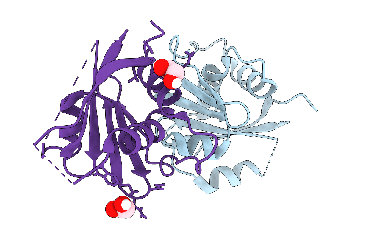
Crystal structure of the N-domain of HMA6, a copper-transporting P-type ATPase
Biological Source:
Source Organism:
Host Organism:
Method Details:
Experimental Method:
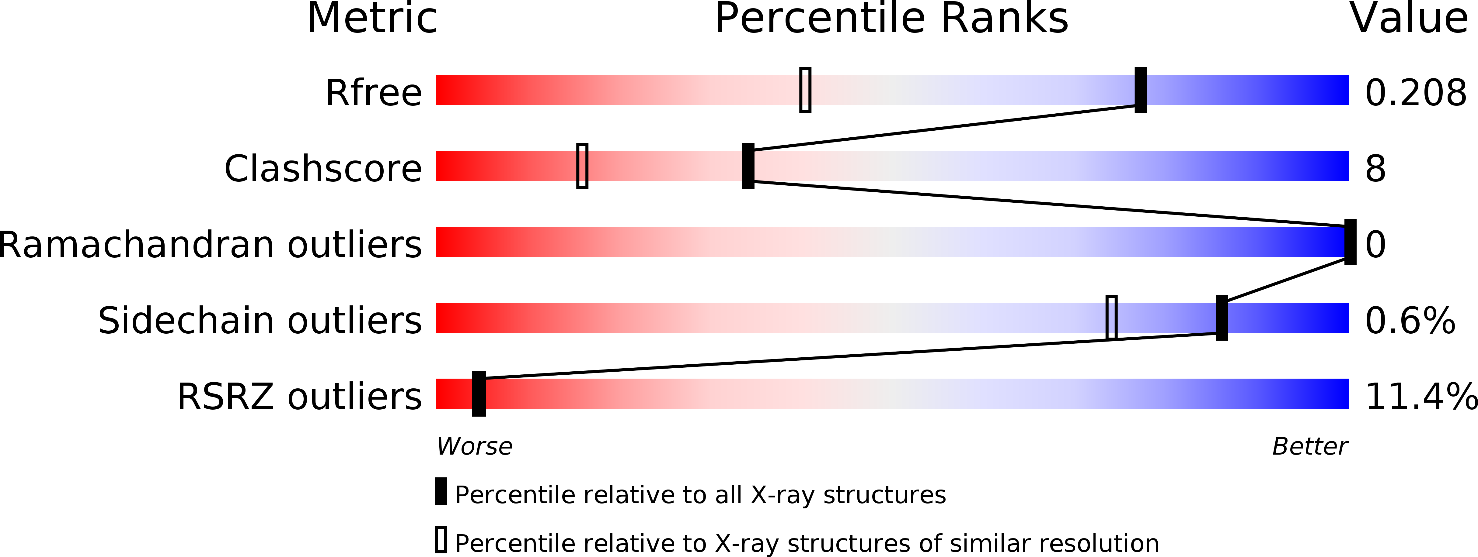
Resolution:
1.50 Å
R-Value Free:
0.20
R-Value Work:
0.18
R-Value Observed:
0.18
Space Group:
P 2 21 21
Macromolecular Entities
Polymer Type:polypeptide(L)
Molecule:Copper-transporting ATPase PAA1, chloroplastic
Gene (Uniprot):PAA1
Mutations:EE709AA
Chain IDs:A, B
Chain Length:131
Number of Molecules:2
Biological Source:Arabidopsis thaliana
Modified Residue
Compound ID Chain ID Parent Comp ID Details 2D Image
MSE A MET modified residue
Primary Citation
Structural Insights into the Nucleotide-Binding Domains of the P1B-type ATPases HMA6 and HMA8 from Arabidopsis thaliana.
PLoS ONE 11 e0165666 e0165666 (2016)
PMID: 27802305 DOI: 10.1371/journal.pone.0165666

Abstact

Copper is a crucial ion in cells, but needs to be closely controlled due to its toxic potential and ability to catalyse the formation of radicals. In chloroplasts, an important step for the proper functioning of the photosynthetic electron transfer chain is the delivery of copper to plastocyanin in the thylakoid lumen. The main route for copper transport to the thylakoid lumen is driven by two PIB-type ATPases, Heavy Metal ATPase 6 (HMA6) and HMA8, located in the inner membrane of the chloroplast envelope and in the thylakoid membrane, respectively. Here, the crystal structures of the nucleotide binding domain of HMA6 and HMA8 from Arabidopsis thaliana are reported at 1.5Å and 1.75Å resolution, respectively, providing the first structural information on plants Cu+-ATPases. The structures reveal a compact domain, with two short helices on both sides of a twisted beta-sheet. A double mutant, aiding in the crystallization, provides a new crystal contact, but also avoids an internal clash highlighting the benefits of construct modifications. Finally, the histidine in the HP motif of the isolated domains, unable to bind ATP, shows a side chain conformation distinct from nucleotide bound structures.

Legend

Protein

Chemical

Disease

Primary Citation of related structures