Deposition Date
2016-06-14
Release Date
2017-07-12
Last Version Date
2024-01-10
Entry Detail
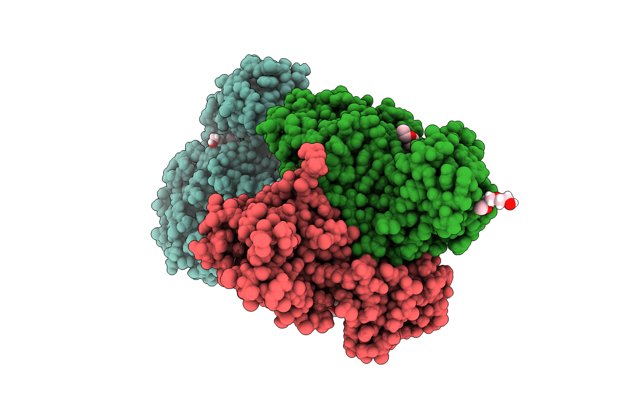
Biological Source:
Source Organism(s):
Cavally virus (Taxon ID: 1041929)
synthetic construct (Taxon ID: 32630)
synthetic construct (Taxon ID: 32630)
Expression System(s):
Method Details:
Experimental Method:
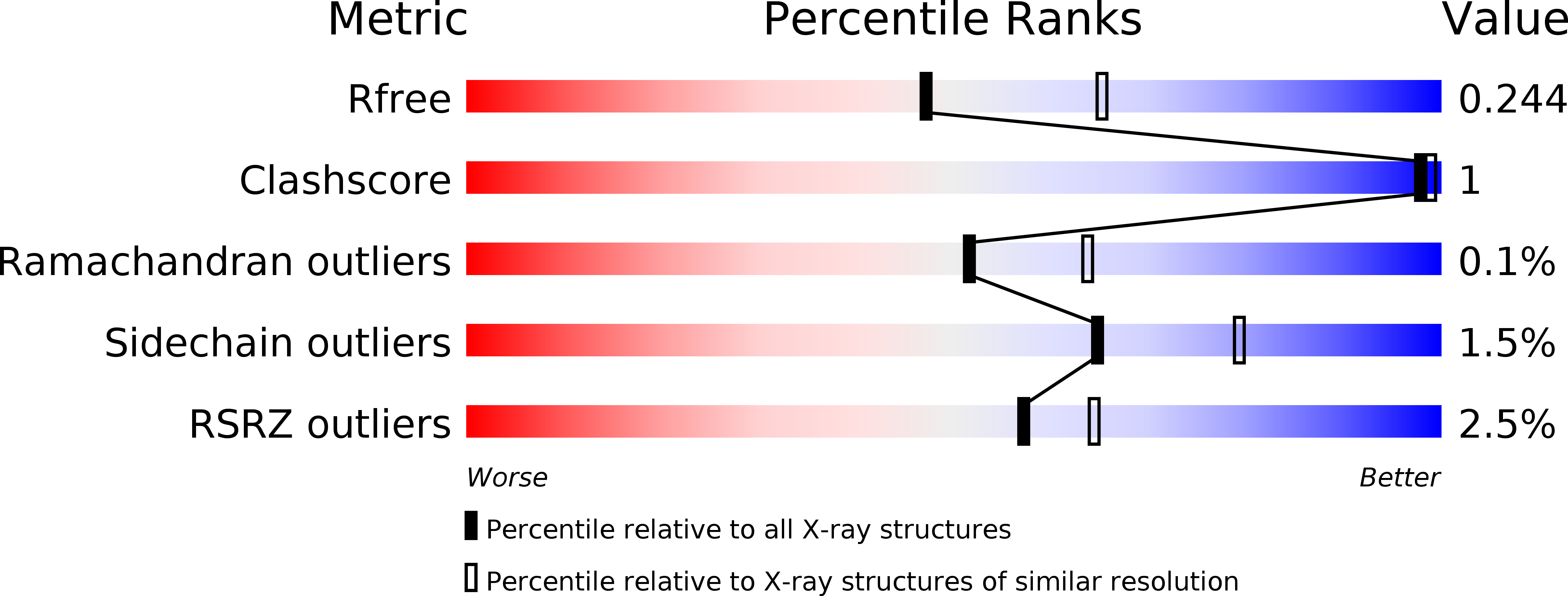
Resolution:
2.30 Å
R-Value Free:
0.22
R-Value Work:
0.20
R-Value Observed:
0.20
Space Group:
P 1 21 1