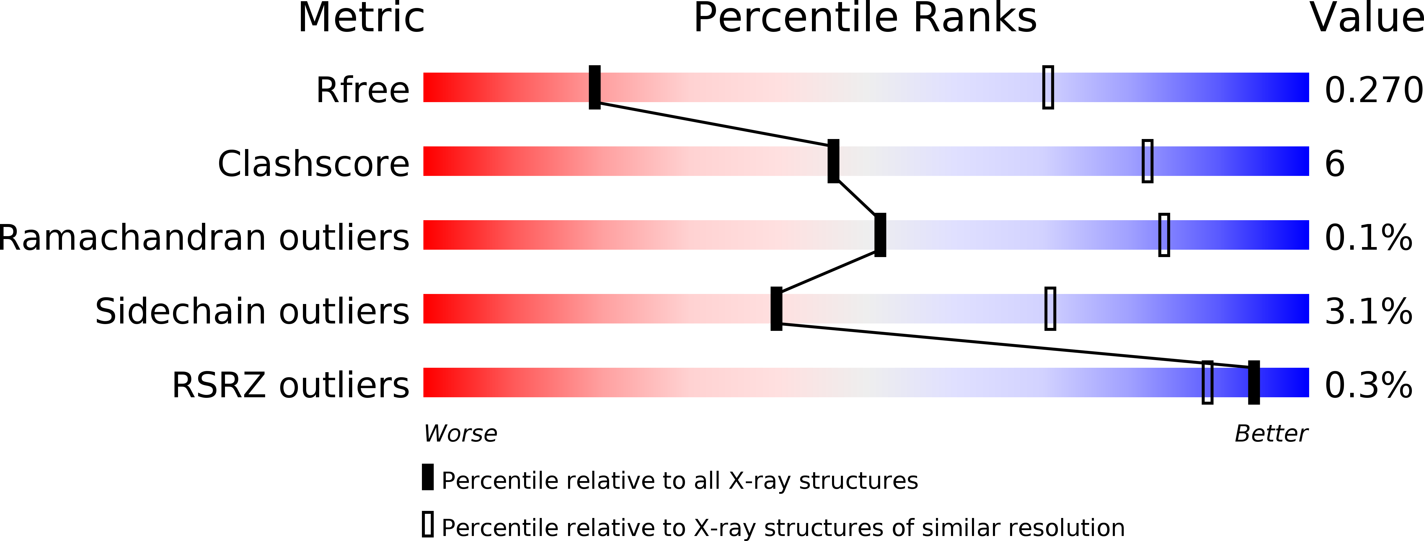
5L7O image
Deposition Date 2016-06-03
Release Date 2016-08-31
Last Version Date 2024-01-10
Entry Detail
PDB ID:
5L7O
Keywords:
Title:
X-ray structure of Triatoma virus empty capsid
Biological Source:
Source Organism:
Method Details:
Experimental Method:
Resolution:
3.60 Å
R-Value Free:
0.26
R-Value Work:
0.26
R-Value Observed:
0.26
Space Group:
H 3
Macromolecular Entities
Structures with similar UniProt ID
Protein Blast
Polymer Type:polypeptide(L)
Molecule:Capsid protein
Chain IDs:A
Chain Length:271
Number of Molecules:1
Biological Source:Triatoma virus
Structures with similar UniProt ID
Protein Blast
Polymer Type:polypeptide(L)
Molecule:Capsid protein
Chain IDs:B
Chain Length:255
Number of Molecules:1
Biological Source:Triatoma virus
Structures with similar UniProt ID
Protein Blast
Polymer Type:polypeptide(L)
Molecule:Capsid protein
Chain IDs:C
Chain Length:285
Number of Molecules:1
Biological Source:Triatoma virus
Ligand Molecules
Primary Citation
X-ray structure of Triatoma virus empty capsid: insights into the mechanism of uncoating and RNA release in dicistroviruses.
J.Gen.Virol. 97 2769 2779 (2016)
PMID: 27519423 DOI: 10.1099/jgv.0.000580

Abstact

In viruses, uncoating and RNA release are two key steps of successfully infecting a target cell. During these steps, the capsid must undergo the necessary conformational changes to allow RNA egress. Despite their importance, these processes are poorly understood in the family Dicistroviridae. Here, we used X-ray crystallography to solve the atomic structure of a Triatoma virus(TrV) empty particle (Protein Data Bank ID 5L7O), which is the resulting capsid after RNA release. It is observed that the overall shape of the capsid and of the three individual proteins is maintained in comparison with the mature virion. Furthermore, no channels indicative of RNA release are formed in the TrV empty particle. However, the most prominent change in the empty particle when compared with the mature virion is the loss of order in the N-terminal domain of the VP2 protein. In mature virions, the VP2 N-terminal domain of one pentamer is swapped with its twofold related copy in an adjacent pentamer, thereby stabilizing the binding between the pentamers. The loss of these interactions allows us to propose that RNA release may take place through transient flipping-out of pentameric subunits. The lower number of stabilizing interactions between the pentamers and the lack of formation of new holes support this model. This model differs from the currently accepted model for rhinoviruses and enteroviruses, in which genome externalization occurs by extrusion of the RNA through capsid channels.

Legend

Protein

Chemical

Disease

Primary Citation of related structures