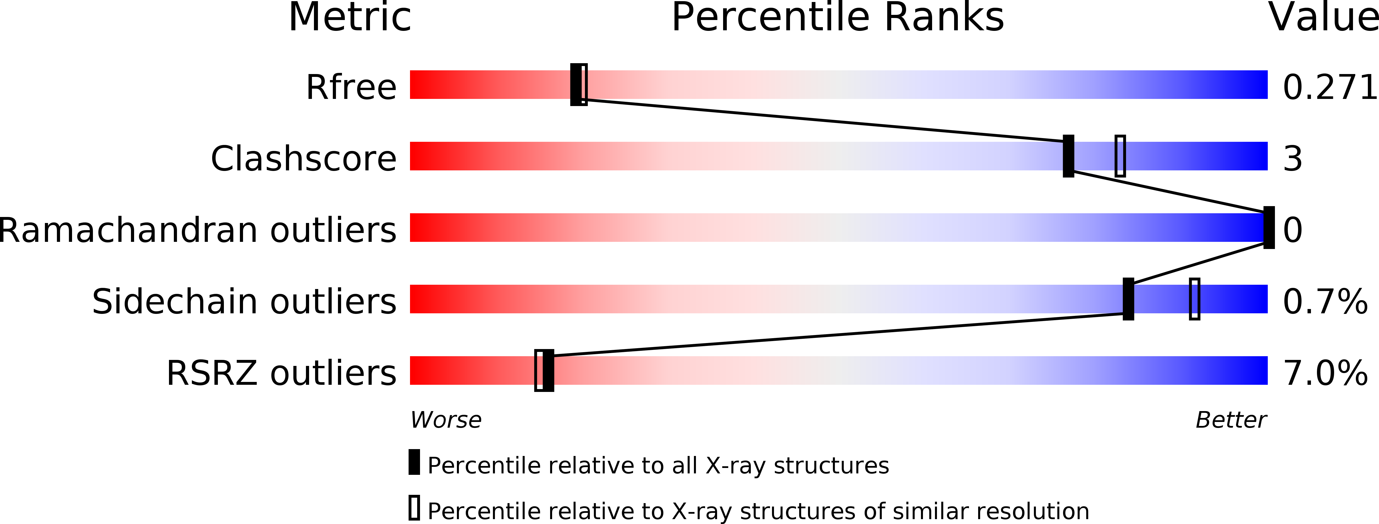
5L7N image
Entry Detail
PDB ID:
5L7N
Title:
Plexin A1 extracellular fragment, domains 7-10 (IPT3-IPT6)
Biological Source:
Source Organism:
Host Organism:
PDB Version:
Deposition Date:
2016-06-03
Release Date:
2017-03-15
Method Details:
Experimental Method:
Resolution:
2.20 Å
R-Value Free:
0.27
R-Value Work:
0.22
R-Value Observed:
0.23
Space Group:
C 2 2 21
Macromolecular Entities
Polymer Type:polypeptide(L)
Description:Plexin-A1
Chain IDs:A
Chain Length:393
Number of Molecules:1
Biological Source:Mus musculus
Primary Citation
Structural Basis for Plexin Activation and Regulation.
Neuron 91 548 560 (2016)
PMID: 27397516 DOI: 10.1016/j.neuron.2016.06.018

Abstact

Class A plexins (PlxnAs) act as semaphorin receptors and control diverse aspects of nervous system development and plasticity, ranging from axon guidance and neuron migration to synaptic organization. PlxnA signaling requires cytoplasmic domain dimerization, but extracellular regulation and activation mechanisms remain unclear. Here we present crystal structures of PlxnA (PlxnA1, PlxnA2, and PlxnA4) full ectodomains. Domains 1-9 form a ring-like conformation from which the C-terminal domain 10 points away. All our PlxnA ectodomain structures show autoinhibitory, intermolecular "head-to-stalk" (domain 1 to domain 4-5) interactions, which are confirmed by biophysical assays, live cell fluorescence microscopy, and cell-based and neuronal growth cone collapse assays. This work reveals a 2-fold role of the PlxnA ectodomains: imposing a pre-signaling autoinhibitory separation for the cytoplasmic domains via intermolecular head-to-stalk interactions and supporting dimerization-based PlxnA activation upon ligand binding. More generally, our data identify a novel molecular mechanism for preventing premature activation of axon guidance receptors.

Legend

Protein

Chemical

Disease

Primary Citation of related structures