
Deposition Date
2016-06-03
Release Date
2016-08-03
Last Version Date
2024-01-10
Entry Detail
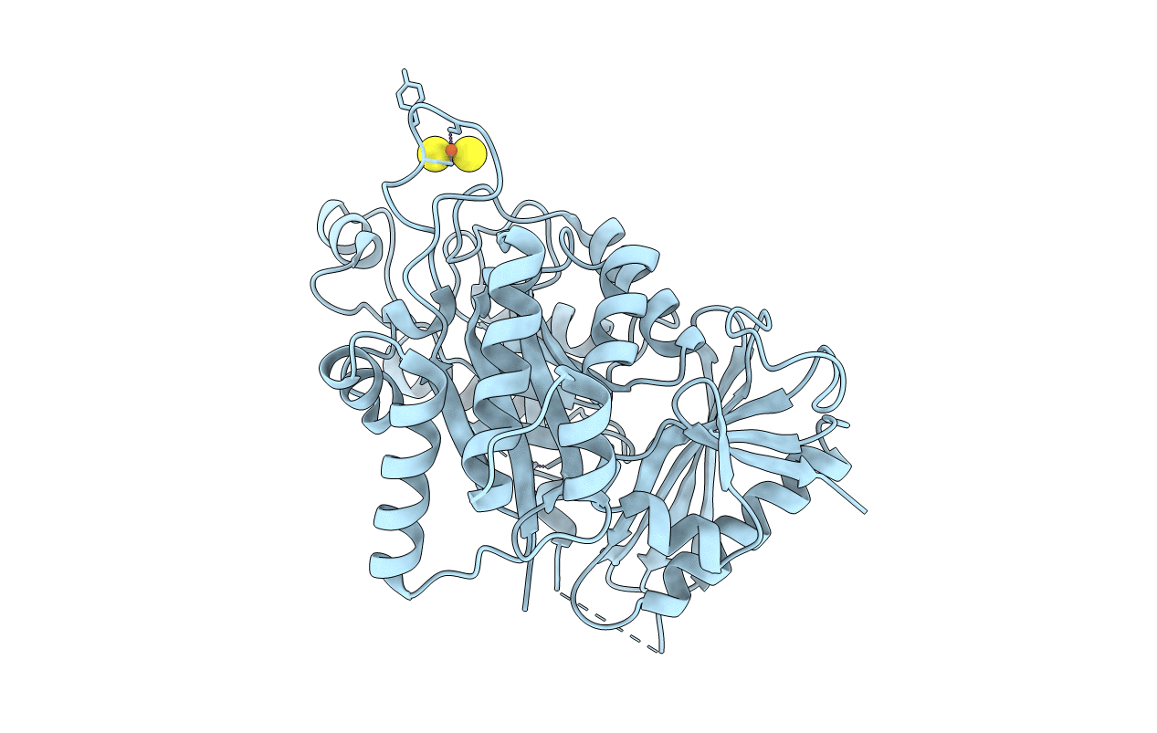
PDB ID:
5L7L
Keywords:
Title:
Crystal Structure of Elp3 from Dehalococcoides mccartyi (390-407 GSGSG)
Biological Source:
Source Organism(s):
Dehalococcoides mccartyi BTF08 (Taxon ID: 1193806)
Expression System(s):
Method Details:
Experimental Method:
Resolution:
2.59 Å
R-Value Free:
0.21
R-Value Work:
0.18
R-Value Observed:
0.18
Space Group:
C 2 2 21
