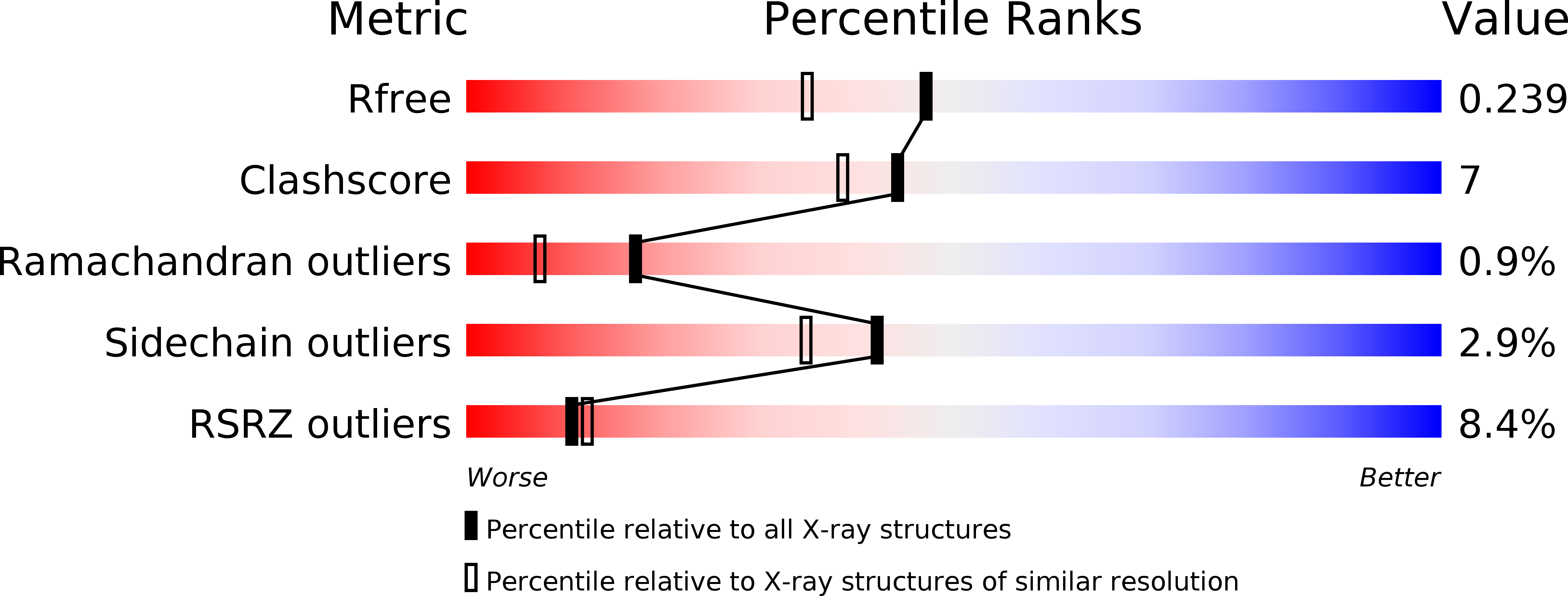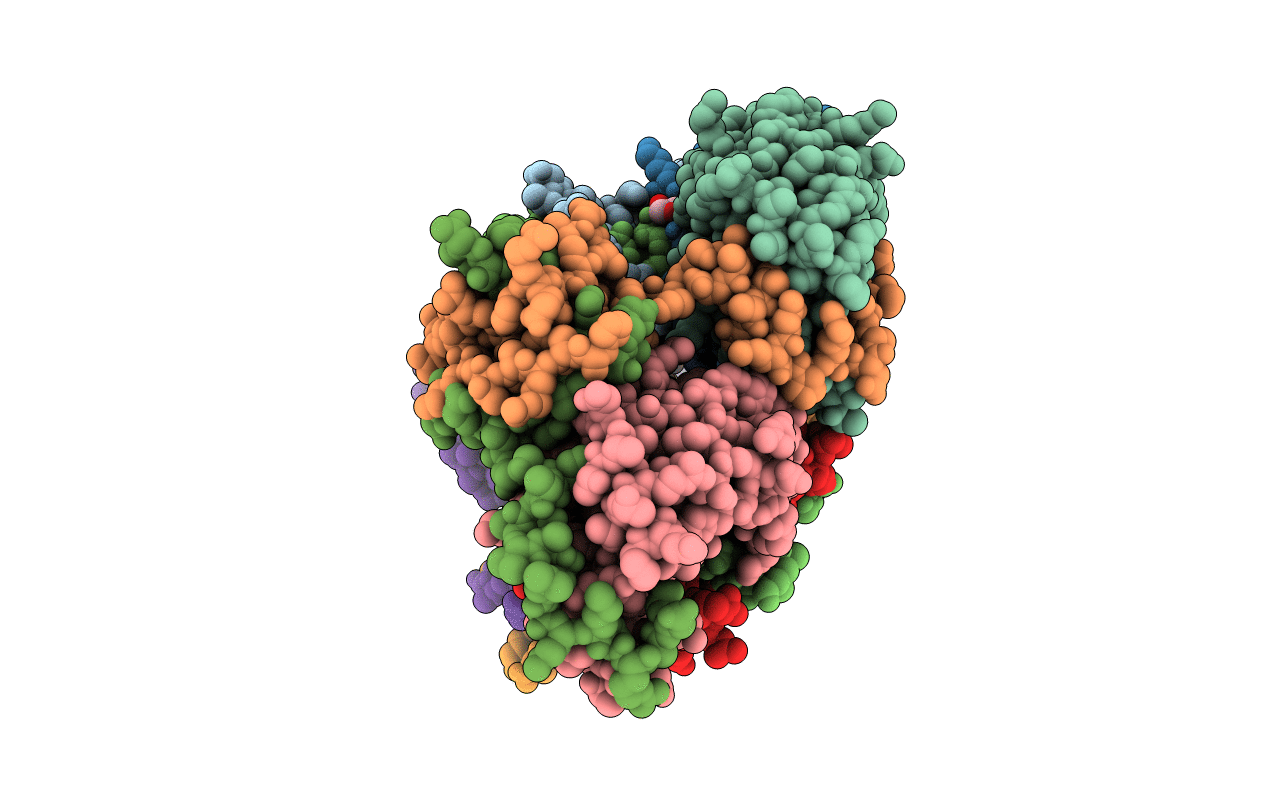
5L6M image
Deposition Date 2016-05-30
Release Date 2016-12-28
Last Version Date 2024-01-10
Entry Detail
PDB ID:
5L6M
Keywords:
Title:
Structure of Caulobacter crescentus VapBC1 (VapB1deltaC:VapC1 form)
Biological Source:
Method Details:
Experimental Method:
Resolution:
1.90 Å
R-Value Free:
0.23
R-Value Work:
0.19
R-Value Observed:
0.19
Space Group:
C 1 2 1
Macromolecular Entities
Polymer Type:polypeptide(L)
Molecule:VapB family protein
Gene (Uniprot):CC_0032
Chain IDs:A, D, E, H, I, L
Chain Length:78
Number of Molecules:6
Biological Source:Caulobacter crescentus (strain ATCC 19089 / CB15)
Polymer Type:polypeptide(L)
Molecule:Ribonuclease VapC
Gene (Uniprot):vapC
Chain IDs:B, C, F, G, J, K
Chain Length:128
Number of Molecules:6
Biological Source:Caulobacter crescentus (strain ATCC 19089 / CB15)
Primary Citation
Toxin inhibition in C. crescentus VapBC1 is mediated by a flexible pseudo-palindromic protein motif and modulated by DNA binding.
Nucleic Acids Res. 45 2875 2886 (2017)
PMID: 27998932 DOI: 10.1093/nar/gkw1266

Abstact

Expression of bacterial type II toxin-antitoxin (TA) systems is regulated at the transcriptional level through direct binding of the antitoxin to pseudo-palindromic sequences on operator DNA. In this context, the toxin functions as a co-repressor by stimulating DNA binding through direct interaction with the antitoxin. Here, we determine crystal structures of the complete 90 kDa heterooctameric VapBC1 complex from Caulobacter crescentus CB15 both in isolation and bound to its cognate DNA operator sequence at 1.6 and 2.7 Å resolution, respectively. DNA binding is associated with a dramatic architectural rearrangement of conserved TA interactions in which C-terminal extended structures of the antitoxin VapB1 swap positions to interlock the complex in the DNA-bound state. We further show that a pseudo-palindromic protein sequence in the antitoxin is responsible for this interaction and required for binding and inactivation of the VapC1 toxin dimer. Sequence analysis of 4127 orthologous VapB sequences reveals that such palindromic protein sequences are widespread and unique to bacterial and archaeal VapB antitoxins suggesting a general principle governing regulation of VapBC TA systems. Finally, a structure of C-terminally truncated VapB1 bound to VapC1 reveals discrete states of the TA interaction that suggest a structural basis for toxin activation in vivo.

Legend

Protein

Chemical

Disease

Primary Citation of related structures