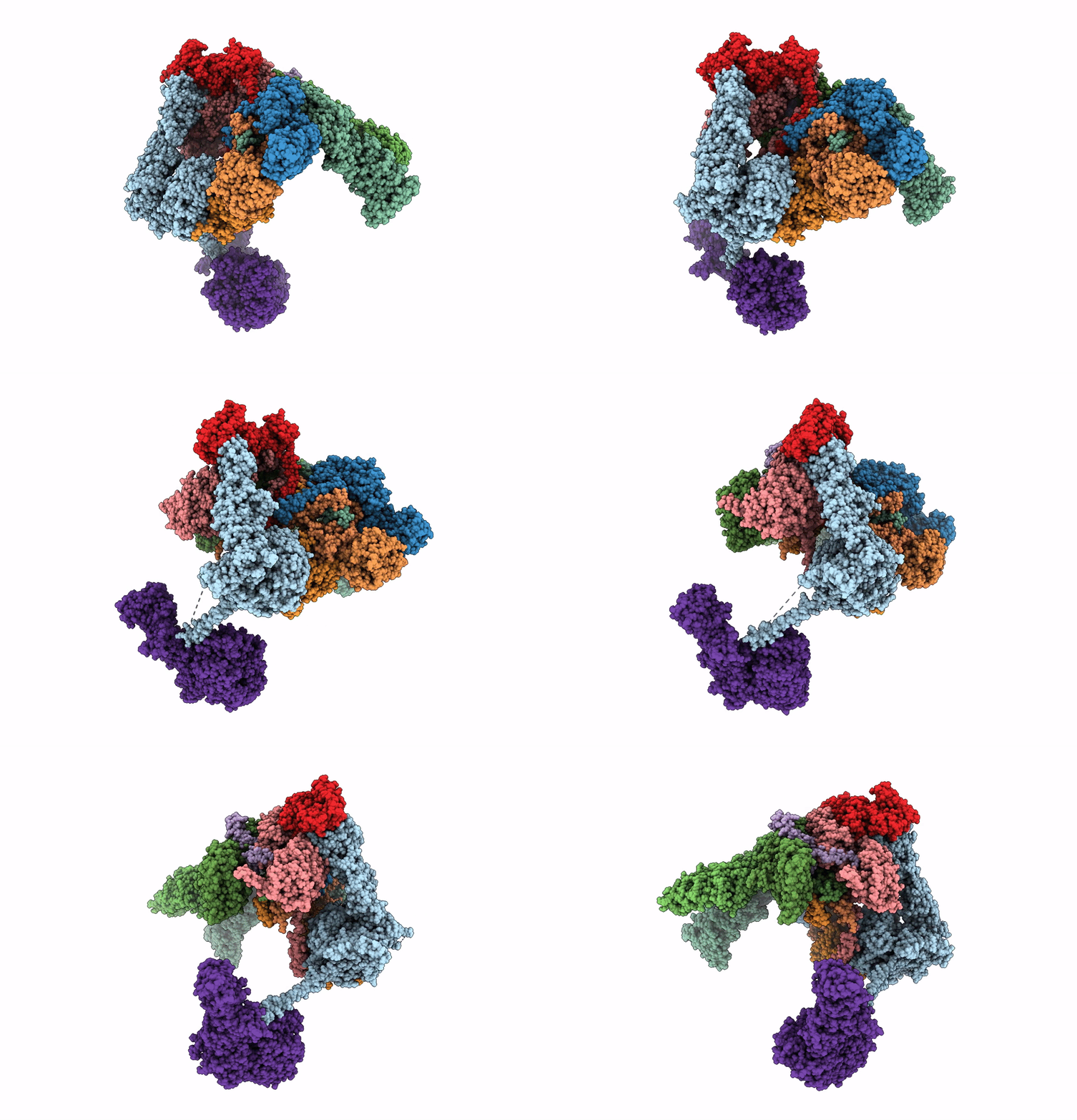
5L4K image
Deposition Date 2016-05-25
Release Date 2016-09-07
Last Version Date 2024-05-08
Entry Detail
PDB ID:
5L4K
Title:
The human 26S proteasome lid
Biological Source:
Source Organism:
Homo sapiens (Taxon ID: 9606)
Method Details:
Experimental Method:
Resolution:
3.90 Å
Aggregation State:
PARTICLE
Reconstruction Method:
SINGLE PARTICLE
Macromolecular Entities
Structures with similar UniProt ID
Protein Blast
Polymer Type:polypeptide(L)
Molecule:26S proteasome non-ATPase regulatory subunit 1
Gene (Uniprot):PSMD1
Chain IDs:F (auth: N)
Chain Length:953
Number of Molecules:1
Biological Source:Homo sapiens
Structures with similar UniProt ID
Protein Blast
Polymer Type:polypeptide(L)
Molecule:26S proteasome non-ATPase regulatory subunit 13
Gene (Uniprot):PSMD13
Chain IDs:L (auth: O)
Chain Length:376
Number of Molecules:1
Biological Source:Homo sapiens
Structures with similar UniProt ID
Protein Blast
Polymer Type:polypeptide(L)
Molecule:26S proteasome non-ATPase regulatory subunit 12
Gene (Uniprot):PSMD12
Chain IDs:H (auth: P)
Chain Length:456
Number of Molecules:1
Biological Source:Homo sapiens
Structures with similar UniProt ID
Protein Blast
Polymer Type:polypeptide(L)
Molecule:26S proteasome non-ATPase regulatory subunit 11
Gene (Uniprot):PSMD11
Chain IDs:I (auth: Q)
Chain Length:422
Number of Molecules:1
Biological Source:Homo sapiens
Structures with similar UniProt ID
Protein Blast
Polymer Type:polypeptide(L)
Molecule:26S proteasome non-ATPase regulatory subunit 6
Gene (Uniprot):PSMD6
Chain IDs:J (auth: R)
Chain Length:389
Number of Molecules:1
Biological Source:Homo sapiens
Structures with similar UniProt ID
Protein Blast
Polymer Type:polypeptide(L)
Molecule:26S proteasome non-ATPase regulatory subunit 3
Gene (Uniprot):PSMD3
Chain IDs:G (auth: S)
Chain Length:534
Number of Molecules:1
Biological Source:Homo sapiens
Structures with similar UniProt ID
Protein Blast
Polymer Type:polypeptide(L)
Molecule:26S proteasome non-ATPase regulatory subunit 8
Gene (Uniprot):PSMD8
Chain IDs:C (auth: T)
Chain Length:350
Number of Molecules:1
Biological Source:Homo sapiens
Structures with similar UniProt ID
Protein Blast
Polymer Type:polypeptide(L)
Molecule:26S proteasome non-ATPase regulatory subunit 7
Gene (Uniprot):PSMD7
Chain IDs:K (auth: U)
Chain Length:324
Number of Molecules:1
Biological Source:Homo sapiens
Structures with similar UniProt ID
Protein Blast
Polymer Type:polypeptide(L)
Molecule:26S proteasome non-ATPase regulatory subunit 14
Gene (Uniprot):PSMD14
Chain IDs:B (auth: V)
Chain Length:310
Number of Molecules:1
Biological Source:Homo sapiens
Structures with similar UniProt ID
Protein Blast
Polymer Type:polypeptide(L)
Molecule:26S proteasome non-ATPase regulatory subunit 4
Gene (Uniprot):PSMD4
Chain IDs:A (auth: W)
Chain Length:377
Number of Molecules:1
Biological Source:Homo sapiens
Structures with similar UniProt ID
Protein Blast
Polymer Type:polypeptide(L)
Molecule:26S proteasome complex subunit DSS1
Gene (Uniprot):SEM1
Chain IDs:D (auth: Y)
Chain Length:70
Number of Molecules:1
Biological Source:Homo sapiens
Structures with similar UniProt ID
Protein Blast
Polymer Type:polypeptide(L)
Molecule:26S proteasome non-ATPase regulatory subunit 2
Gene (Uniprot):PSMD2
Chain IDs:E (auth: Z)
Chain Length:908
Number of Molecules:1
Biological Source:Homo sapiens
Ligand Molecules
Primary Citation
Structure of the human 26S proteasome at a resolution of 3.9 angstrom.
Proc.Natl.Acad.Sci.USA 113 7816 7821 (2016)
PMID: 27342858 DOI: 10.1073/pnas.1608050113

Abstact

Protein degradation in eukaryotic cells is performed by the Ubiquitin-Proteasome System (UPS). The 26S proteasome holocomplex consists of a core particle (CP) that proteolytically degrades polyubiquitylated proteins, and a regulatory particle (RP) containing the AAA-ATPase module. This module controls access to the proteolytic chamber inside the CP and is surrounded by non-ATPase subunits (Rpns) that recognize substrates and deubiquitylate them before unfolding and degradation. The architecture of the 26S holocomplex is highly conserved between yeast and humans. The structure of the human 26S holocomplex described here reveals previously unidentified features of the AAA-ATPase heterohexamer. One subunit, Rpt6, has ADP bound, whereas the other five have ATP in their binding pockets. Rpt6 is structurally distinct from the other five Rpt subunits, most notably in its pore loop region. For Rpns, the map reveals two main, previously undetected, features: the C terminus of Rpn3 protrudes into the mouth of the ATPase ring; and Rpn1 and Rpn2, the largest proteasome subunits, are linked by an extended connection. The structural features of the 26S proteasome observed in this study are likely to be important for coordinating the proteasomal subunits during substrate processing.

Legend

Protein

Chemical

Disease

Primary Citation of related structures