5L46 image
Deposition Date 2016-05-25
Release Date 2016-08-17
Last Version Date 2024-11-20
Entry Detail
PDB ID:
5L46
Keywords:
Title:
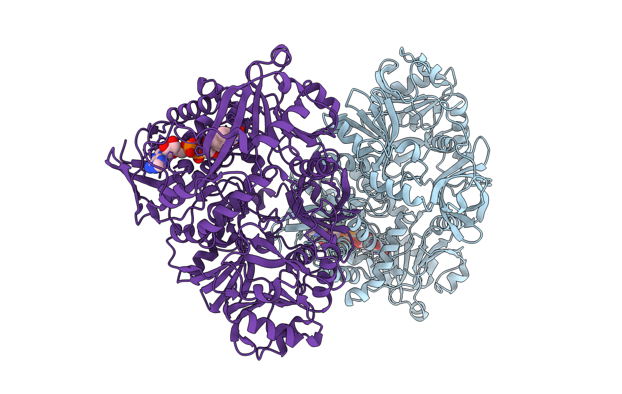
Crystal structure of human dimethylglycine-dehydrogenase
Biological Source:
Source Organism:
Homo sapiens (Taxon ID: 9606)
Host Organism:
Method Details:
Experimental Method:
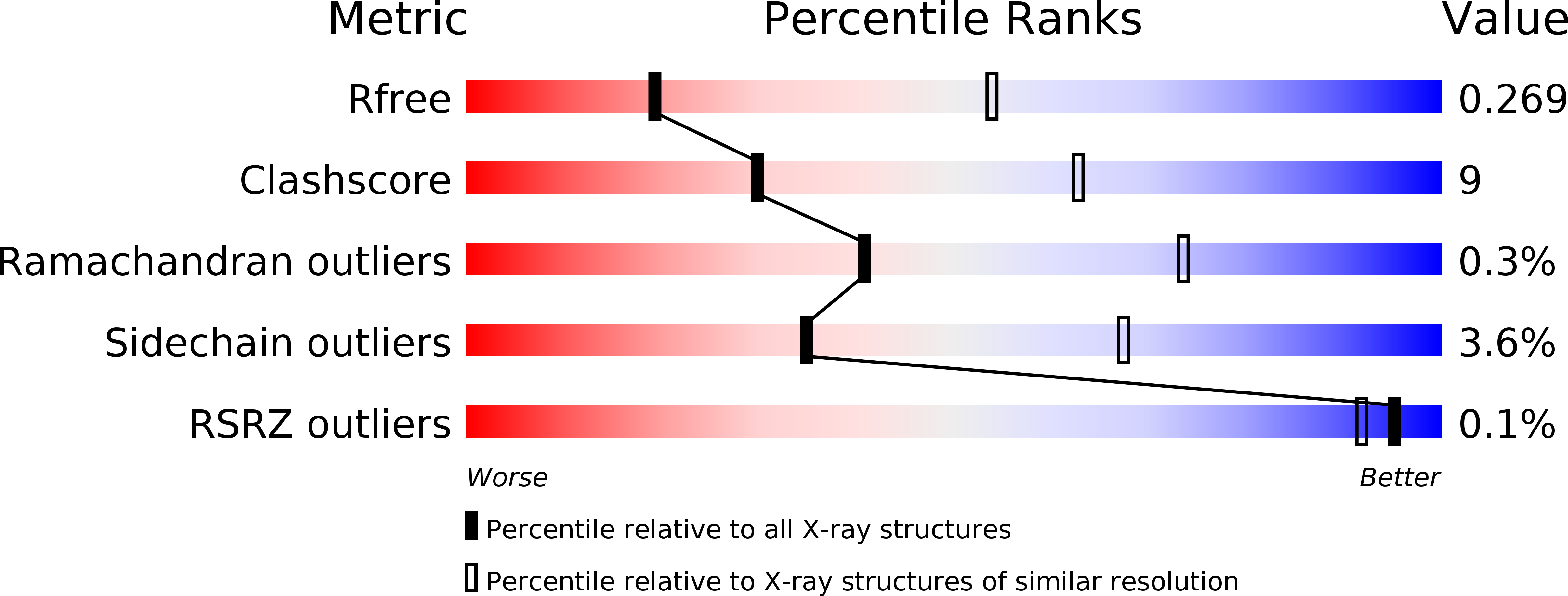
Resolution:
3.09 Å
R-Value Free:
0.26
R-Value Work:
0.17
R-Value Observed:
0.18
Space Group:
P 1 21 1
Macromolecular Entities
Polymer Type:polypeptide(L)
Molecule:Dimethylglycine dehydrogenase, mitochondrial
Gene (Uniprot):DMGDH
Chain IDs:A, B
Chain Length:841
Number of Molecules:2
Biological Source:Homo sapiens
Ligand Molecules
Primary Citation
Structure and biochemical properties of recombinant human dimethylglycine dehydrogenase and comparison to the disease-related H109R variant.
Febs J. 283 3587 3603 (2016)
PMID: 27486859 DOI: 10.1111/febs.13828

Abstact

UNLABELLED The human dimethylglycine dehydrogenase (hDMGDH) is a flavin adenine dinucleotide (FAD)- and tetrahydrofolate (THF)-dependent, mitochondrial matrix enzyme taking part in choline degradation, one-carbon metabolism and electron transfer to the respiratory chain. The rare natural variant H109R causes dimethylglycine dehydrogenase deficiency leading to increased blood and urinary dimethylglycine concentrations. A detailed biochemical and structural characterization of hDMGDH was thus far hampered by insufficient heterologous expression of the protein. In the present study, we report the development of an intracellular, heterologous expression system in Komagataella phaffii (formerly known as Pichia pastoris) providing the opportunity to determine kinetic parameters, spectroscopic properties, thermostability, and the redox potential of hDMGDH. Moreover, we have successfully crystallized the wild-type enzyme and determined the structure to 3.1-Å resolution. The structure-based analysis of our biochemical data provided new insights into the kinetic properties of the enzyme in particular with respect to oxygen reactivity. A comparative study with the H109R variant demonstrated that the variant suffers from decreased protein stability, cofactor saturation, and substrate affinity. DATABASE Structural data are available in the PDB database under the accession number 5L46.

Legend

Protein

Chemical

Disease

Primary Citation of related structures