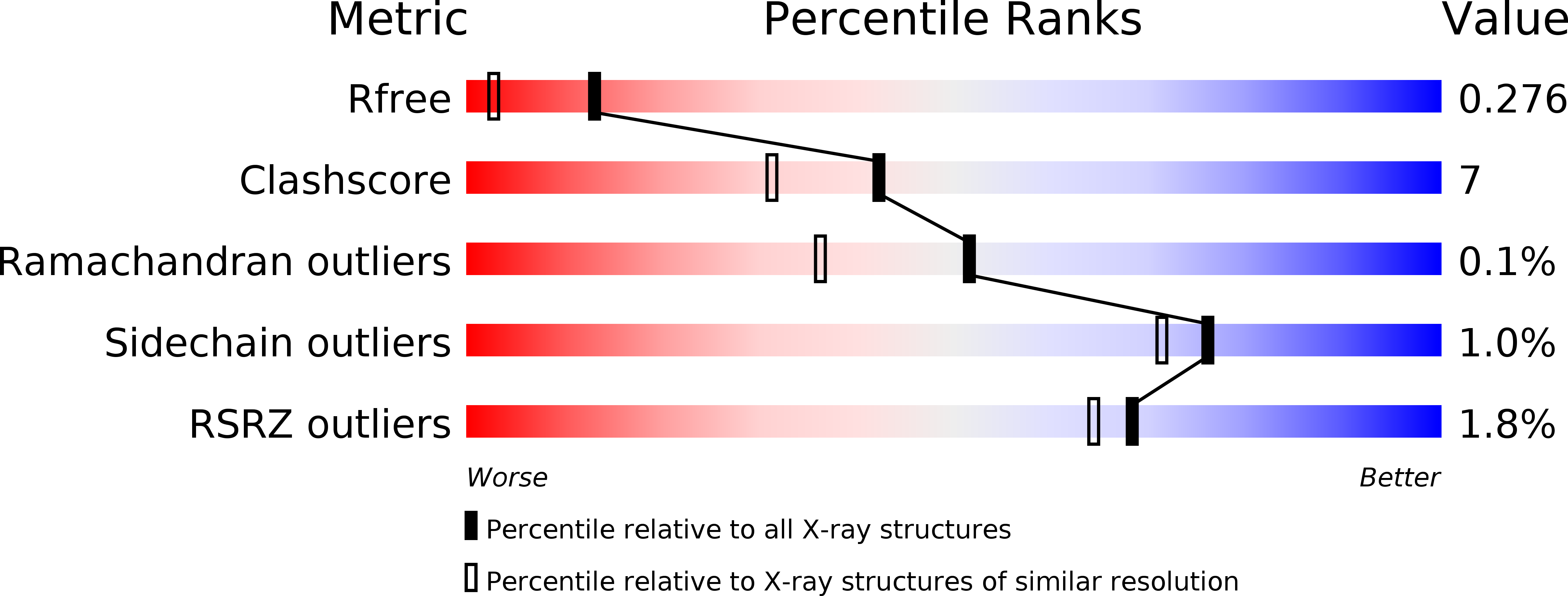
5KYO image
Entry Detail
PDB ID:
5KYO
Keywords:
Title:
Crystal Structure of CYP101J2
Biological Source:
Source Organism:
Host Organism:
PDB Version:
Deposition Date:
2016-07-22
Release Date:
2017-01-04
Method Details:
Experimental Method:
Resolution:
1.80 Å
R-Value Free:
0.27
R-Value Work:
0.22
R-Value Observed:
0.22
Space Group:
P 1
Macromolecular Entities
Polymer Type:polypeptide(L)
Description:CYP101J2
Chain IDs:A, B, C, D, E, F
Chain Length:430
Number of Molecules:6
Biological Source:Sphingobium yanoikuyae
Ligand Molecules
Primary Citation
X-ray crystal structure of cytochrome P450 monooxygenase CYP101J2 from Sphingobium yanoikuyae strain B2.
Proteins 85 945 950 (2017)
PMID: 27936485 DOI: 10.1002/prot.25227

Abstact

The cytochrome P450 monooxygenases (P450s) catalyze a vast array of oxygenation reactions that can be useful in biocatalytic applications. CYP101J2 from Sphingobium yanoikuyae is a P450 that catalyzes the hydroxylation of 1,8-cineole. Here we report the crystallization and X-ray structure elucidation of recombinant CYP101J2 to 1.8 Å resolution. The CYP101J2 structure shows the canonical P450-fold and has an open conformation in the absence of substrate. Analysis of the structure revealed that CYP101J2, in the absence of substrate, forms a well-ordered substrate-binding channel that suggests a unique form of substrate guidance in comparison to other bacterial 1,8-cineole-hydroxylating P450 enzymes. Proteins 2017; 85:945-950. © 2016 Wiley Periodicals, Inc.

Legend

Protein

Chemical

Disease

Primary Citation of related structures