5KW9 image
Deposition Date 2016-07-15
Release Date 2016-10-05
Last Version Date 2024-11-13
Entry Detail
PDB ID:
5KW9
Title:
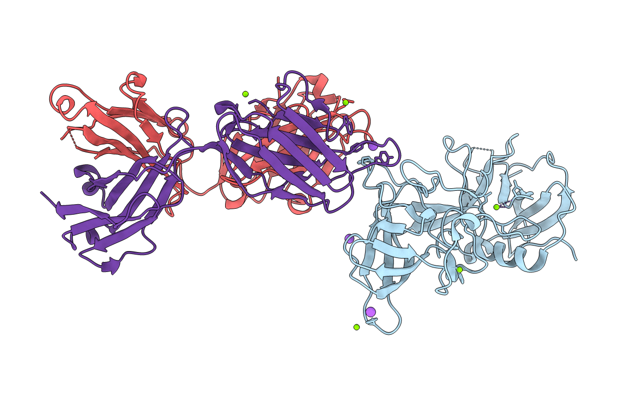
Structural Basis for Norovirus Neutralization by a HBGA Blocking Human IgA Antibody
Biological Source:
Source Organism:
Norwalk virus (Taxon ID: 11983)
Homo sapiens (Taxon ID: 9606)
Host Organism:
Method Details:
Experimental Method:
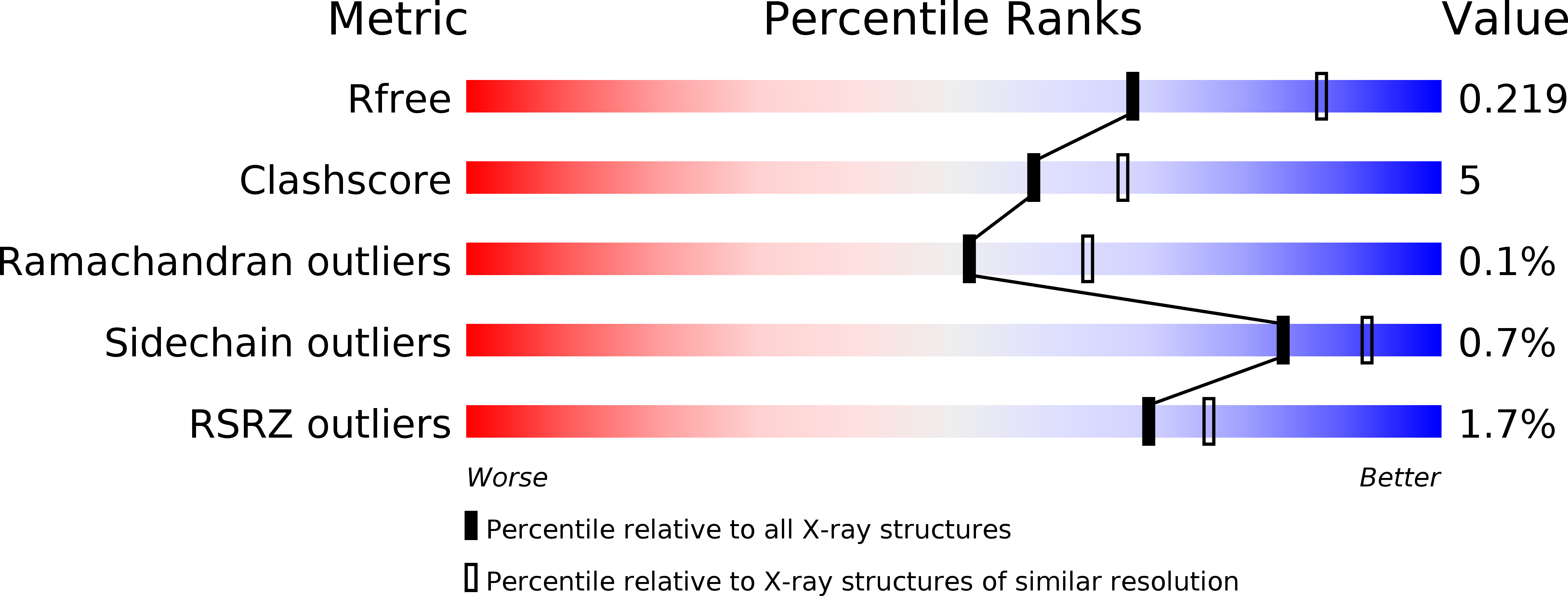
Resolution:
2.30 Å
R-Value Free:
0.21
R-Value Work:
0.18
R-Value Observed:
0.18
Space Group:
P 65 2 2
Macromolecular Entities
Polymer Type:polypeptide(L)
Molecule:Capsid protein VP1
Gene (Uniprot):ORF2
Chain IDs:A
Chain Length:295
Number of Molecules:1
Biological Source:Norwalk virus
Polymer Type:polypeptide(L)
Molecule:IgA(VH)-IgG(CH) heavy chain Fab fragment
Chain IDs:B (auth: H)
Chain Length:227
Number of Molecules:1
Biological Source:Homo sapiens
Polymer Type:polypeptide(L)
Molecule:IgA Light chain
Chain IDs:C (auth: L)
Chain Length:220
Number of Molecules:1
Biological Source:Homo sapiens
Primary Citation
Structural basis for norovirus neutralization by an HBGA blocking human IgA antibody.
Proc.Natl.Acad.Sci.USA 113 E5830 E5837 (2016)
PMID: 27647885 DOI: 10.1073/pnas.1609990113

Abstact

Human noroviruses (HuNoVs) cause sporadic and epidemic gastroenteritis worldwide. They are classified into two major genogroups (GI and GII), with each genogroup further divided into multiple genotypes. Susceptibility to these viruses is influenced by genetically determined histo-blood group antigen (HBGA) expression. HBGAs function as cell attachment factors by binding to a surface-exposed region in the protruding (P) domain of the capsid protein. Sequence variations in this region that result in differential HBGA binding patterns and antigenicity are suggested to form a basis for strain diversification. Recent studies show that serum antibodies that block HBGA binding correlate with protection against illness. Although genogroup-dependent variation in HBGA binding specificity is structurally well characterized, an understanding of how antibodies block HBGA binding and how genotypic variations affect such blockade is lacking. Our crystallographic studies of the GI.1 P domain in complex with the Fab fragment of a human IgA monoclonal antibody (IgA 5I2) with HBGA blocking activity show that the antibody recognizes a conformational epitope formed by two surface-exposed loop clusters in the P domain. The antibody engulfs the HBGA binding site but does not affect its structural integrity. An unusual feature of the antigen recognition by IgA 5I2 is the predominant involvement of the CDR light chain 1 in contrast to the commonly observed CDR heavy chain 3, providing a unique perspective into antibody diversity in antigen recognition. Identification of the antigenic site in the P domain shows how genotypic variations might allow escape from antibody neutralization and exemplifies the interplay between antigenicity and HBGA specificity in HuNoV evolution.

Legend

Protein

Chemical

Disease

Primary Citation of related structures