5KUG image
Deposition Date 2016-07-13
Release Date 2016-09-07
Last Version Date 2023-10-04
Entry Detail
PDB ID:
5KUG
Keywords:
Title:
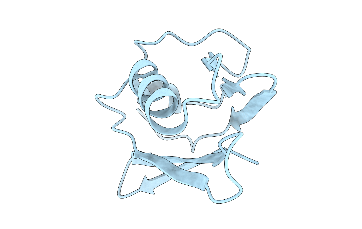
Human mitochondrial calcium uniporter (residues 72-189) crystal structure with lithium
Biological Source:
Source Organism:
Homo sapiens (Taxon ID: 9606)
Host Organism:
Method Details:
Experimental Method:
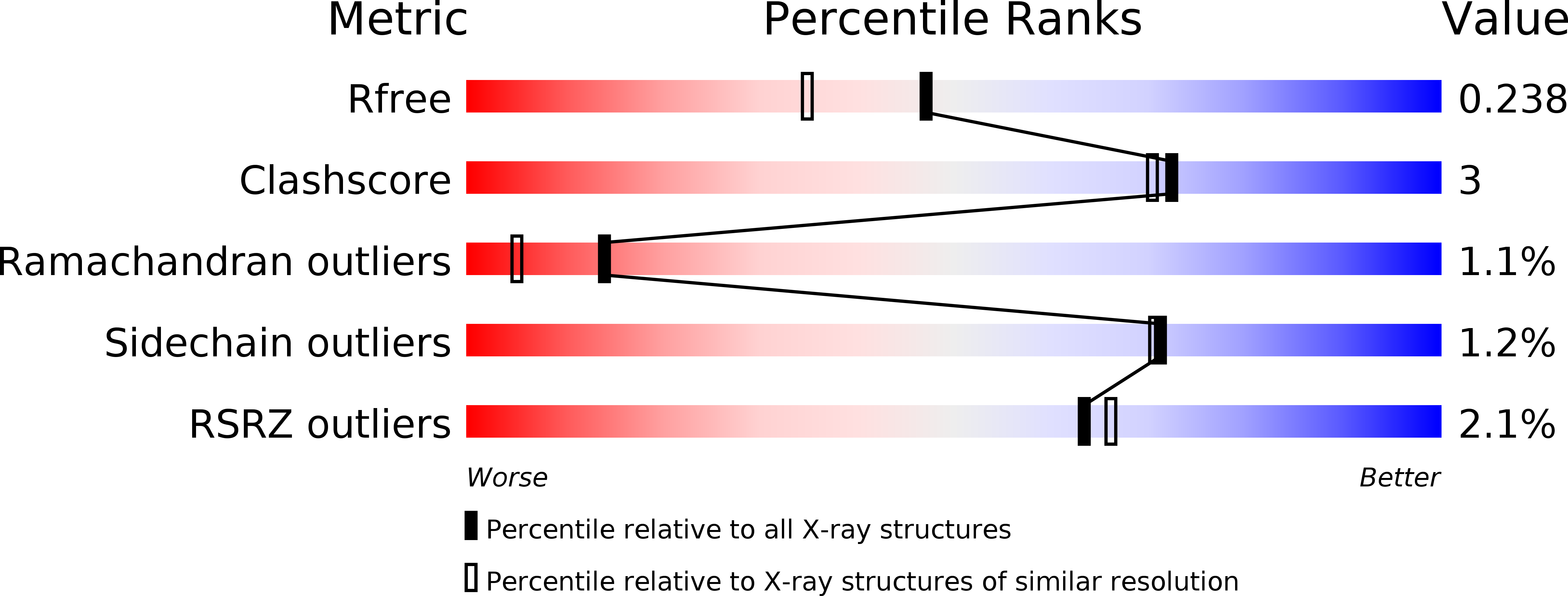
Resolution:
1.90 Å
R-Value Free:
0.23
R-Value Work:
0.20
R-Value Observed:
0.20
Space Group:
P 65
Macromolecular Entities
Polymer Type:polypeptide(L)
Molecule:Calcium uniporter protein, mitochondrial
Gene (Uniprot):MCU
Chain IDs:A
Chain Length:124
Number of Molecules:1
Biological Source:Homo sapiens
Primary Citation
Structural Insights into Mitochondrial Calcium Uniporter Regulation by Divalent Cations.
Cell Chem Biol 23 1157 1169 (2016)
PMID: 27569754 DOI: 10.1016/j.chembiol.2016.07.012

Abstact

Calcium (Ca(2+)) flux into the matrix is tightly controlled by the mitochondrial Ca(2+) uniporter (MCU) due to vital roles in cell death and bioenergetics. However, the precise atomic mechanisms of MCU regulation remain unclear. Here, we solved the crystal structure of the N-terminal matrix domain of human MCU, revealing a β-grasp-like fold with a cluster of negatively charged residues that interacts with divalent cations. Binding of Ca(2+) or Mg(2+) destabilizes and shifts the self-association equilibrium of the domain toward monomer. Mutational disruption of the acidic face weakens oligomerization of the isolated matrix domain and full-length human protein similar to cation binding and markedly decreases MCU activity. Moreover, mitochondrial Mg(2+) loading or blockade of mitochondrial Ca(2+) extrusion suppresses MCU Ca(2+)-uptake rates. Collectively, our data reveal that the β-grasp-like matrix region harbors an MCU-regulating acidic patch that inhibits human MCU activity in response to Mg(2+) and Ca(2+) binding.

Legend

Protein

Chemical

Disease

Primary Citation of related structures