5KNL image
Entry Detail
PDB ID:
5KNL
Keywords:
Title:
Crystal structure of S. pombe ubiquitin E1 (Uba1) in complex with Ubc15 and ubiquitin
Biological Source:
PDB Version:
Deposition Date:
2016-06-28
Release Date:
2017-02-15
Method Details:
Experimental Method:
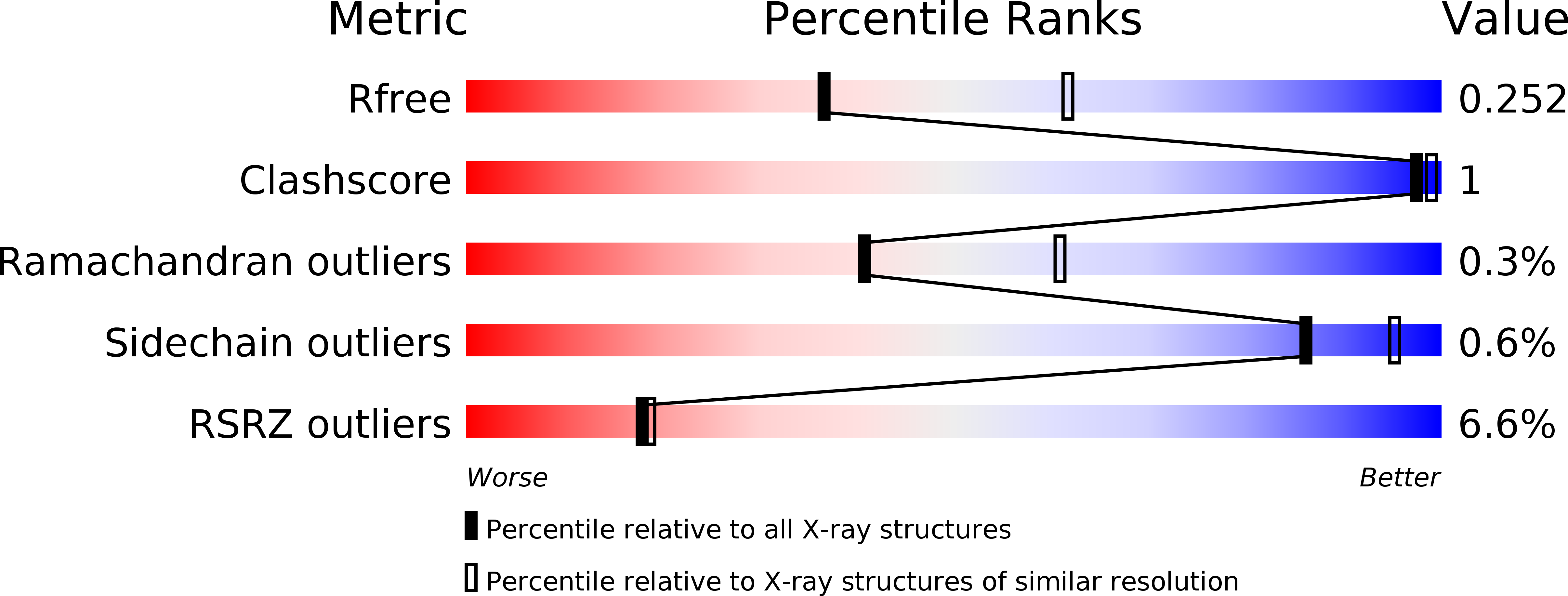
Resolution:
2.50 Å
R-Value Free:
0.25
R-Value Work:
0.21
R-Value Observed:
0.21
Space Group:
P 1
Macromolecular Entities
Polymer Type:polypeptide(L)
Description:Ubiquitin-activating enzyme E1 1
Chain IDs:A, D
Chain Length:1001
Number of Molecules:2
Biological Source:Schizosaccharomyces pombe (strain 972 / ATCC 24843)
Polymer Type:polypeptide(L)
Description:Ubiquitin
Mutations:K6R, K11R, K27R, S28A, K29R, K33R, K48R, S57A, K63R
Chain IDs:B, E
Chain Length:96
Number of Molecules:2
Biological Source:Pseudozyma antarctica
Polymer Type:polypeptide(L)
Description:Ubiquitin-conjugating enzyme E2 15
Chain IDs:C, F
Chain Length:175
Number of Molecules:2
Biological Source:Schizosaccharomyces pombe (strain 972 / ATCC 24843)
Ligand Molecules
Primary Citation
S. pombe Uba1-Ubc15 Structure Reveals a Novel Regulatory Mechanism of Ubiquitin E2 Activity.
Mol. Cell 65 699 714.e6 (2017)
PMID: 28162934 DOI: 10.1016/j.molcel.2017.01.008

Abstact

Ubiquitin (Ub) E1 initiates the Ub conjugation cascade by activating and transferring Ub to tens of different E2s. How Ub E1 cooperates with E2s that differ substantially in their predicted E1-interacting residues is unknown. Here, we report the structure of S. pombe Uba1 in complex with Ubc15, a Ub E2 with intrinsically low E1-E2 Ub thioester transfer activity. The structure reveals a distinct Ubc15 binding mode that substantially alters the network of interactions at the E1-E2 interface compared to the only other available Ub E1-E2 structure. Structure-function analysis reveals that the intrinsically low activity of Ubc15 largely results from the presence of an acidic residue at its N-terminal region. Notably, Ub E2 N termini are serine/threonine rich in many other Ub E2s, leading us to hypothesize that phosphorylation of these sites may serve as a novel negative regulatory mechanism of Ub E2 activity, which we demonstrate biochemically and in cell-based assays.

Legend

Protein

Chemical

Disease

Primary Citation of related structures