Deposition Date
2016-06-27
Release Date
2017-02-08
Last Version Date
2023-09-27
Entry Detail
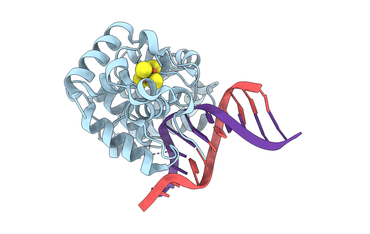
Biological Source:
Source Organism(s):
Geobacillus stearothermophilus (Taxon ID: 1422)
Expression System(s):
Method Details:
Experimental Method:
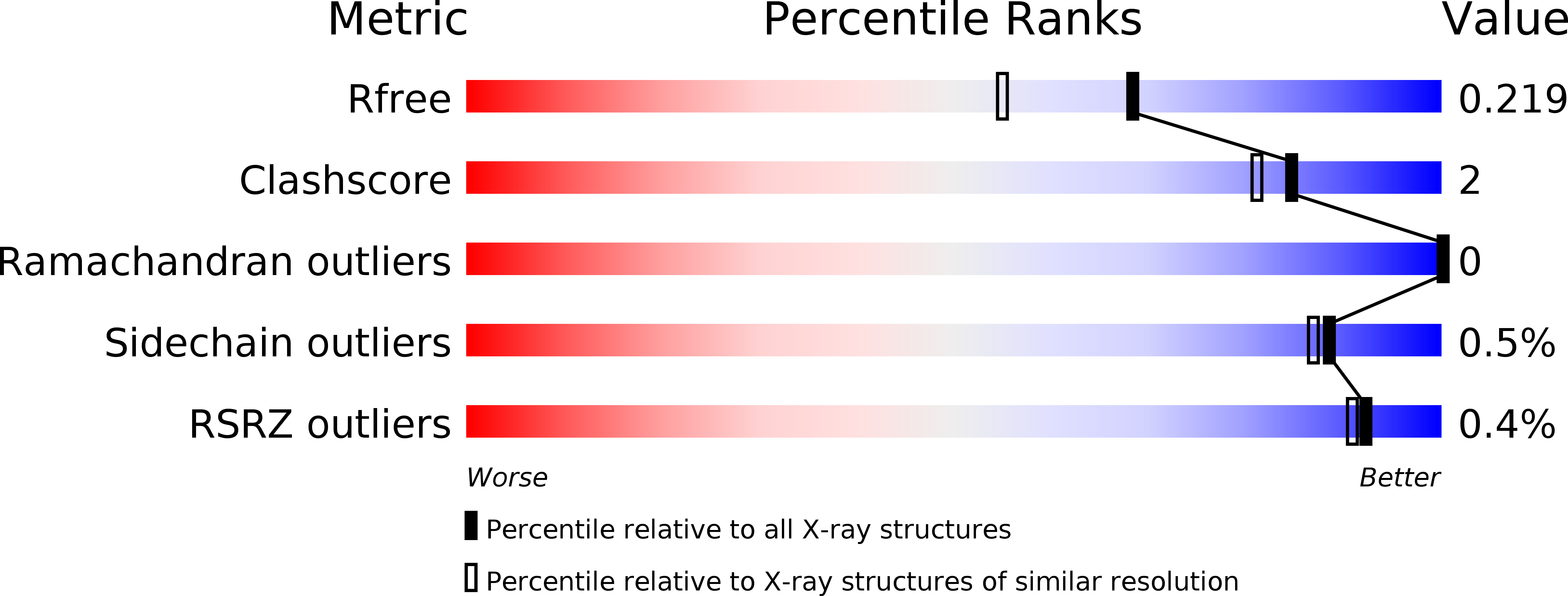
Resolution:
1.81 Å
R-Value Free:
0.21
R-Value Work:
0.16
R-Value Observed:
0.16
Space Group:
P 1 21 1