5KKK image
Entry Detail
PDB ID:
5KKK
Title:
1.7-Angstrom In situ Mylar structure of sperm whale myoglobin (SWMb-CO) at 100 K
Biological Source:
Source Organism:
Host Organism:
PDB Version:
Deposition Date:
2016-06-21
Release Date:
2017-02-15
Method Details:
Experimental Method:
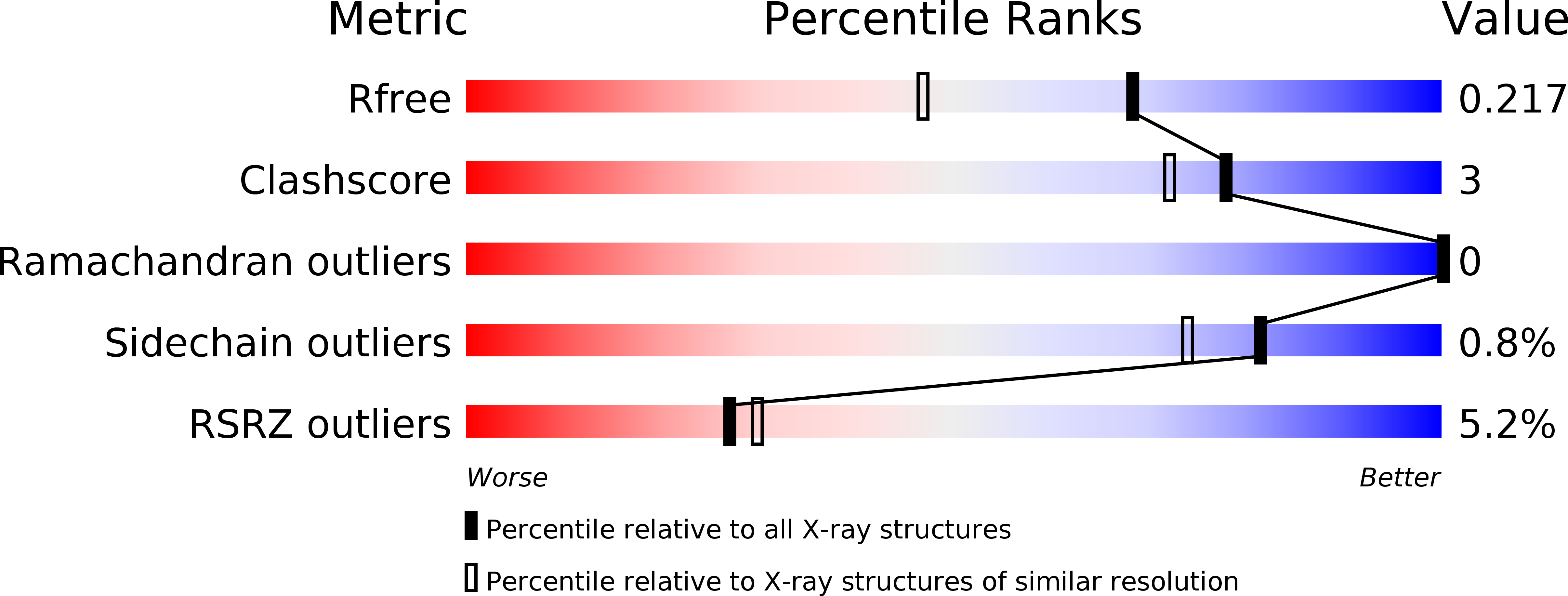
Resolution:
1.70 Å
R-Value Free:
0.21
R-Value Work:
0.17
R-Value Observed:
0.17
Space Group:
P 6
Macromolecular Entities
Polymer Type:polypeptide(L)
Description:Myoglobin
Chain IDs:A
Chain Length:154
Number of Molecules:1
Biological Source:Physeter catodon
Primary Citation
A Versatile System for High-Throughput In Situ X-ray Screening and Data Collection of Soluble and Membrane-Protein Crystals.
Cryst Growth Des 16 6318 6326 (2016)
PMID: 28261000 DOI: 10.1021/acs.cgd.6b00950

Abstact

In recent years, in situ data collection has been a major focus of progress in protein crystallography. Here, we introduce the Mylar in situ method using Mylar-based sandwich plates that are inexpensive, easy to make and handle, and show significantly less background scattering than other setups. A variety of cognate holders for patches of Mylar in situ sandwich films corresponding to one or more wells makes the method robust and versatile, allows for storage and shipping of entire wells, and enables automated crystal imaging, screening, and goniometer-based X-ray diffraction data-collection at room temperature and under cryogenic conditions for soluble and membrane-protein crystals grown in or transferred to these plates. We validated the Mylar in situ method using crystals of the water-soluble proteins hen egg-white lysozyme and sperm whale myoglobin as well as the 7-transmembrane protein bacteriorhodopsin from Haloquadratum walsbyi. In conjunction with current developments at synchrotrons, this approach promises high-resolution structural studies of membrane proteins to become faster and more routine.

Legend

Protein

Chemical

Disease

Primary Citation of related structures