
Deposition Date
2016-06-21
Release Date
2017-03-22
Last Version Date
2024-11-20
Entry Detail
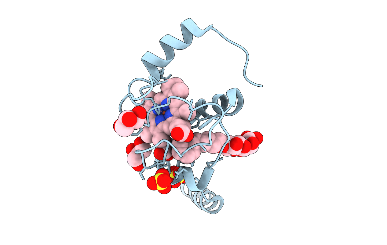
PDB ID:
5KKE
Keywords:
Title:
Crystal Structure of a Domain-swapped Dimer of Yeast Iso-1-cytochrome c with CYMAL5
Biological Source:
Source Organism(s):
Expression System(s):
Method Details:
Experimental Method:
Resolution:
1.70 Å
R-Value Free:
0.20
R-Value Work:
0.17
R-Value Observed:
0.17
Space Group:
I 2 2 2
