5KJ7 image
Deposition Date 2016-06-17
Release Date 2016-10-19
Last Version Date 2023-09-27
Entry Detail
PDB ID:
5KJ7
Title:
Structure of the Ca2+-bound synaptotagmin-1 SNARE complex (long unit cell form) - from XFEL diffraction
Biological Source:
Source Organism:
Host Organism:
Method Details:
Experimental Method:
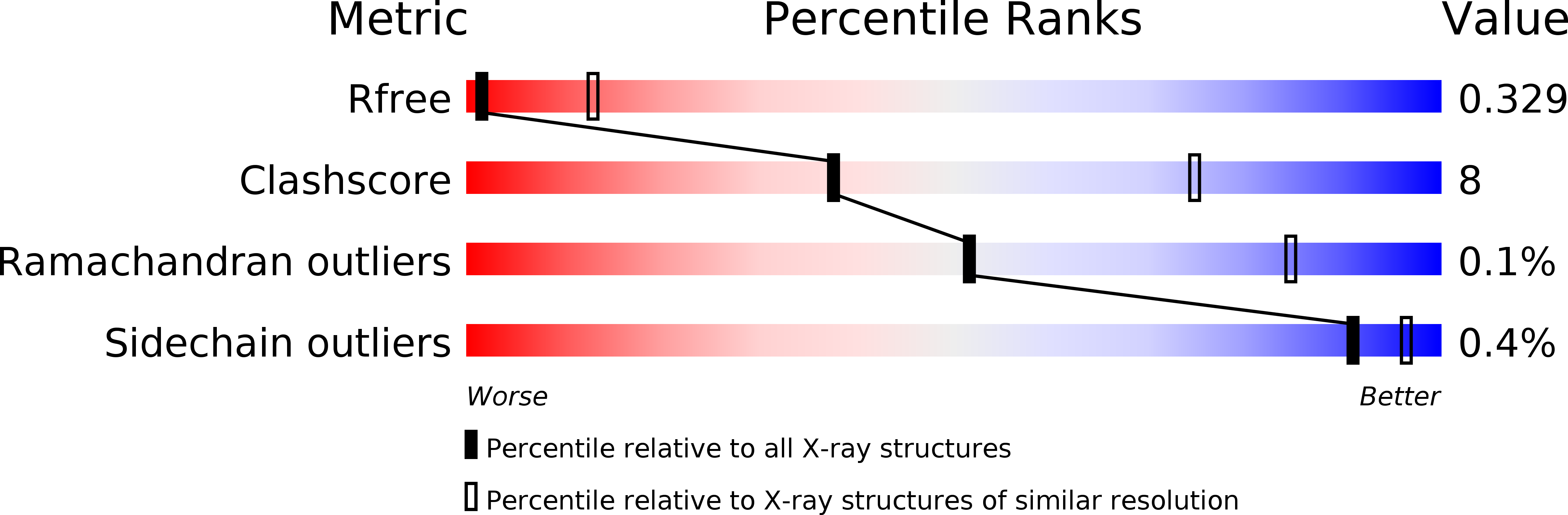
Resolution:
3.50 Å
R-Value Free:
0.32
R-Value Work:
0.29
R-Value Observed:
0.29
Space Group:
P 21 21 21
Macromolecular Entities
Structures with similar UniProt ID
Protein Blast
Polymer Type:polypeptide(L)
Molecule:Vesicle-associated membrane protein 3
Gene (Uniprot):Vamp3
Chain IDs:A, G
Chain Length:63
Number of Molecules:2
Biological Source:Rattus norvegicus
Structures with similar UniProt ID
Protein Blast
Polymer Type:polypeptide(L)
Molecule:Syntaxin-1A
Gene (Uniprot):Stx1a
Chain IDs:B, H
Chain Length:66
Number of Molecules:2
Biological Source:Rattus norvegicus
Structures with similar UniProt ID
Protein Blast
Polymer Type:polypeptide(L)
Molecule:Synaptosomal-associated protein 25
Gene (Uniprot):Snap25
Chain IDs:C, I
Chain Length:75
Number of Molecules:2
Biological Source:Rattus norvegicus
Structures with similar UniProt ID
Protein Blast
Polymer Type:polypeptide(L)
Molecule:Synaptosomal-associated protein 25
Gene (Uniprot):Snap25
Chain IDs:D, J
Chain Length:64
Number of Molecules:2
Biological Source:Rattus norvegicus
Structures with similar UniProt ID
Protein Blast
Polymer Type:polypeptide(L)
Molecule:Synaptotagmin-1
Gene (Uniprot):Syt1
Chain IDs:E, F, K
Chain Length:279
Number of Molecules:3
Biological Source:Rattus norvegicus
Ligand Molecules
Primary Citation
Advances in X-ray free electron laser (XFEL) diffraction data processing applied to the crystal structure of the synaptotagmin-1 / SNARE complex.
Elife 5 ? ? (2016)
PMID: 27731796 DOI: 10.7554/eLife.18740

Abstact

X-ray free electron lasers (XFELs) reduce the effects of radiation damage on macromolecular diffraction data and thereby extend the limiting resolution. Previously, we adapted classical post-refinement techniques to XFEL diffraction data to produce accurate diffraction data sets from a limited number of diffraction images (Uervirojnangkoorn et al., 2015), and went on to use these techniques to obtain a complete data set from crystals of the synaptotagmin-1 / SNARE complex and to determine the structure at 3.5 Å resolution (Zhou et al., 2015). Here, we describe new advances in our methods and present a reprocessed XFEL data set of the synaptotagmin-1 / SNARE complex. The reprocessing produced small improvements in electron density maps and the refined atomic model. The maps also contained more information than those of a lower resolution (4.1 Å) synchrotron data set. Processing a set of simulated XFEL diffraction images revealed that our methods yield accurate data and atomic models.

Legend

Protein

Chemical

Disease

Primary Citation of related structures
Feedback Form
Name
Email
Institute
Feedback