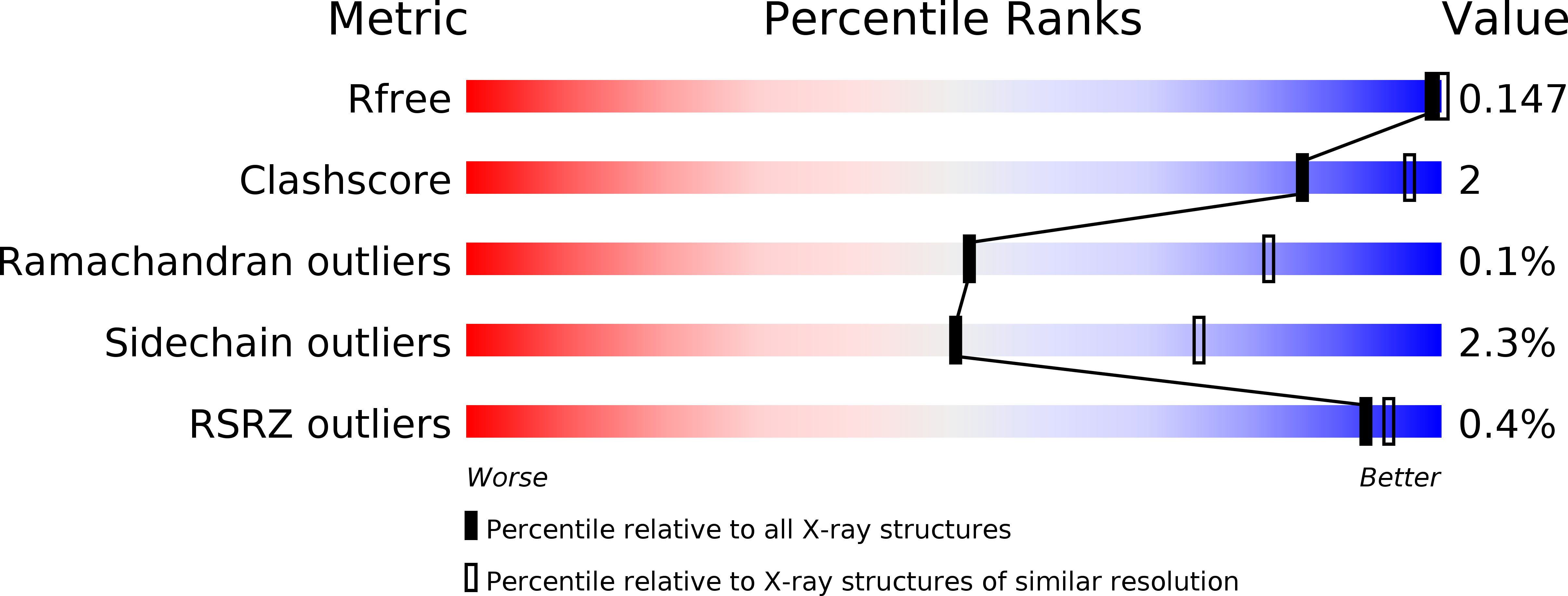
5KJ4 image
Deposition Date 2016-06-17
Release Date 2016-11-23
Last Version Date 2023-09-27
Entry Detail
PDB ID:
5KJ4
Title:
Crystal Structure of Mouse Protocadherin-15 EC9-10
Biological Source:
Source Organism:
Mus musculus (Taxon ID: 10090)
Method Details:
Experimental Method:
Resolution:
3.35 Å
R-Value Free:
0.19
R-Value Work:
0.15
R-Value Observed:
0.15
Space Group:
P 31
Macromolecular Entities
Polymer Type:polypeptide(L)
Molecule:Protocadherin-15
Gene (Uniprot):Pcdh15
Chain IDs:A, B, C, D
Chain Length:234
Number of Molecules:4
Biological Source:Mus musculus
Ligand Molecules
Primary Citation
An elastic element in the protocadherin-15 tip link of the inner ear.
Nat Commun 7 13458 13458 (2016)
PMID: 27857071 DOI: 10.1038/ncomms13458

Abstact

Tip link filaments convey force and gate inner-ear hair-cell transduction channels to mediate perception of sound and head movements. Cadherin-23 and protocadherin-15 form tip links through a calcium-dependent interaction of their extracellular domains made of multiple extracellular cadherin (EC) repeats. These repeats are structurally similar, but not identical in sequence, often featuring linkers with conserved calcium-binding sites that confer mechanical strength to them. Here we present the X-ray crystal structures of human protocadherin-15 EC8-EC10 and mouse EC9-EC10, which show an EC8-9 canonical-like calcium-binding linker, and an EC9-10 calcium-free linker that alters the linear arrangement of EC repeats. Molecular dynamics simulations and small-angle X-ray scattering experiments support this non-linear conformation. Simulations also suggest that unbending of EC9-10 confers some elasticity to otherwise rigid tip links. The new structure provides a first view of protocadherin-15's non-canonical EC linkers and suggests how they may function in inner-ear mechanotransduction, with implications for other cadherins.

Legend

Protein

Chemical

Disease

Primary Citation of related structures