Deposition Date
2016-06-08
Release Date
2016-08-03
Last Version Date
2023-09-27
Entry Detail
PDB ID:
5KDO
Keywords:
Title:
Heterotrimeric complex of the 4 alanine insertion variant of the Gi alpha1 subunit and the Gbeta1-Ggamma1
Biological Source:
Source Organism(s):
Rattus norvegicus (Taxon ID: 10116)
Bos taurus (Taxon ID: 9913)
Bos taurus (Taxon ID: 9913)
Expression System(s):
Method Details:
Experimental Method:
Resolution:
1.90 Å
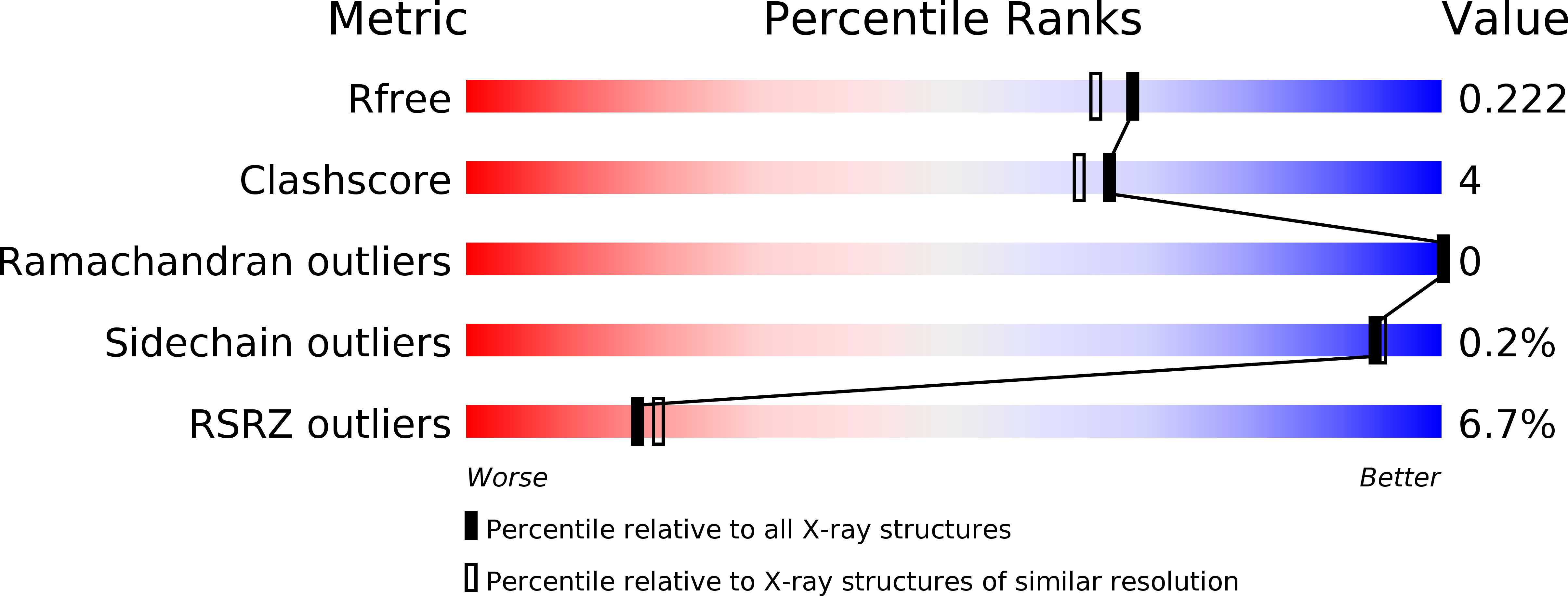
R-Value Free:
0.20
R-Value Work:
0.18
R-Value Observed:
0.18
Space Group:
P 43