5KDL image
Deposition Date 2016-06-08
Release Date 2016-08-03
Last Version Date 2023-09-27
Entry Detail
PDB ID:
5KDL
Title:
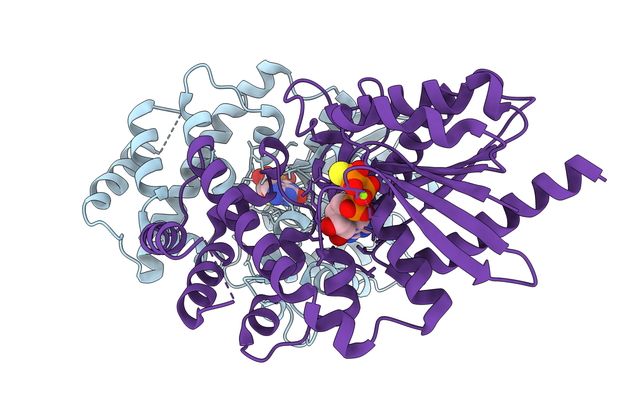
Crystal structure of the 4 alanine insertion variant of the Gi alpha1 subunit bound to GTPgammaS
Biological Source:
Source Organism:
Host Organism:
Method Details:
Experimental Method:
Resolution:
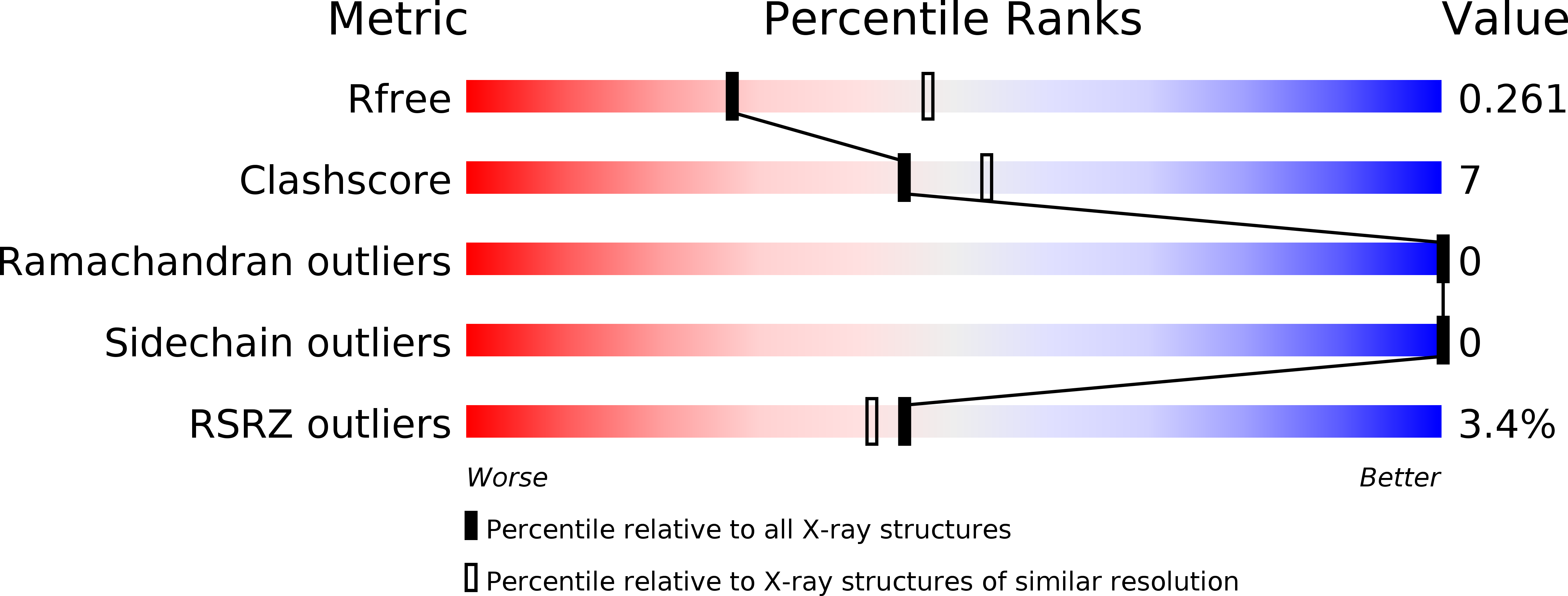
2.67 Å
R-Value Free:
0.26
R-Value Work:
0.20
R-Value Observed:
0.21
Space Group:
P 1 21 1
Macromolecular Entities
Polymer Type:polypeptide(L)
Molecule:Guanine nucleotide-binding protein G(i) subunit alpha-1
Gene (Uniprot):Gnai1
Chain IDs:A, B
Chain Length:358
Number of Molecules:2
Biological Source:Rattus norvegicus
Primary Citation
A Conserved Hydrophobic Core in G alpha i1 Regulates G Protein Activation and Release from Activated Receptor.
J.Biol.Chem. 291 19674 19686 (2016)
PMID: 27462082 DOI: 10.1074/jbc.M116.745513

Abstact

G protein-coupled receptor-mediated heterotrimeric G protein activation is a major mode of signal transduction in the cell. Previously, we and other groups reported that the α5 helix of Gαi1, especially the hydrophobic interactions in this region, plays a key role during nucleotide release and G protein activation. To further investigate the effect of this hydrophobic core, we disrupted it in Gαi1 by inserting 4 alanine amino acids into the α5 helix between residues Gln(333) and Phe(334) (Ins4A). This extends the length of the α5 helix without disturbing the β6-α5 loop interactions. This mutant has high basal nucleotide exchange activity yet no receptor-mediated activation of nucleotide exchange. By using structural approaches, we show that this mutant loses critical hydrophobic interactions, leading to significant rearrangements of side chain residues His(57), Phe(189), Phe(191), and Phe(336); it also disturbs the rotation of the α5 helix and the π-π interaction between His(57) and Phe(189) In addition, the insertion mutant abolishes G protein release from the activated receptor after nucleotide binding. Our biochemical and computational data indicate that the interactions between α5, α1, and β2-β3 are not only vital for GDP release during G protein activation, but they are also necessary for proper GTP binding (or GDP rebinding). Thus, our studies suggest that this hydrophobic interface is critical for accurate rearrangement of the α5 helix for G protein release from the receptor after GTP binding.

Legend

Protein

Chemical

Disease

Primary Citation of related structures