Deposition Date
2016-06-05
Release Date
2016-07-27
Last Version Date
2024-02-07
Entry Detail
PDB ID:
5KC7
Keywords:
Title:
Crystal structure of Cbln1 (Val55-Gly58 deletion mutant)
Biological Source:
Source Organism(s):
Homo sapiens (Taxon ID: 9606)
Expression System(s):
Method Details:
Experimental Method:
Resolution:
7.04 Å
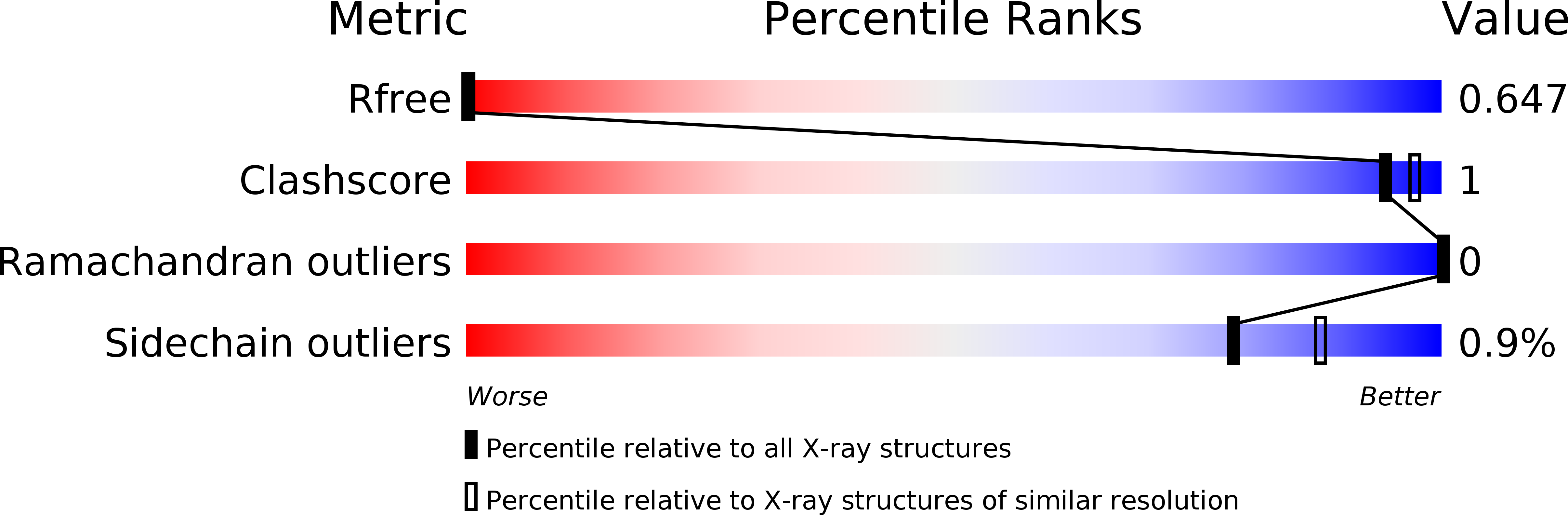
R-Value Free:
0.35
R-Value Work:
0.26
R-Value Observed:
0.27
Space Group:
I 21 3