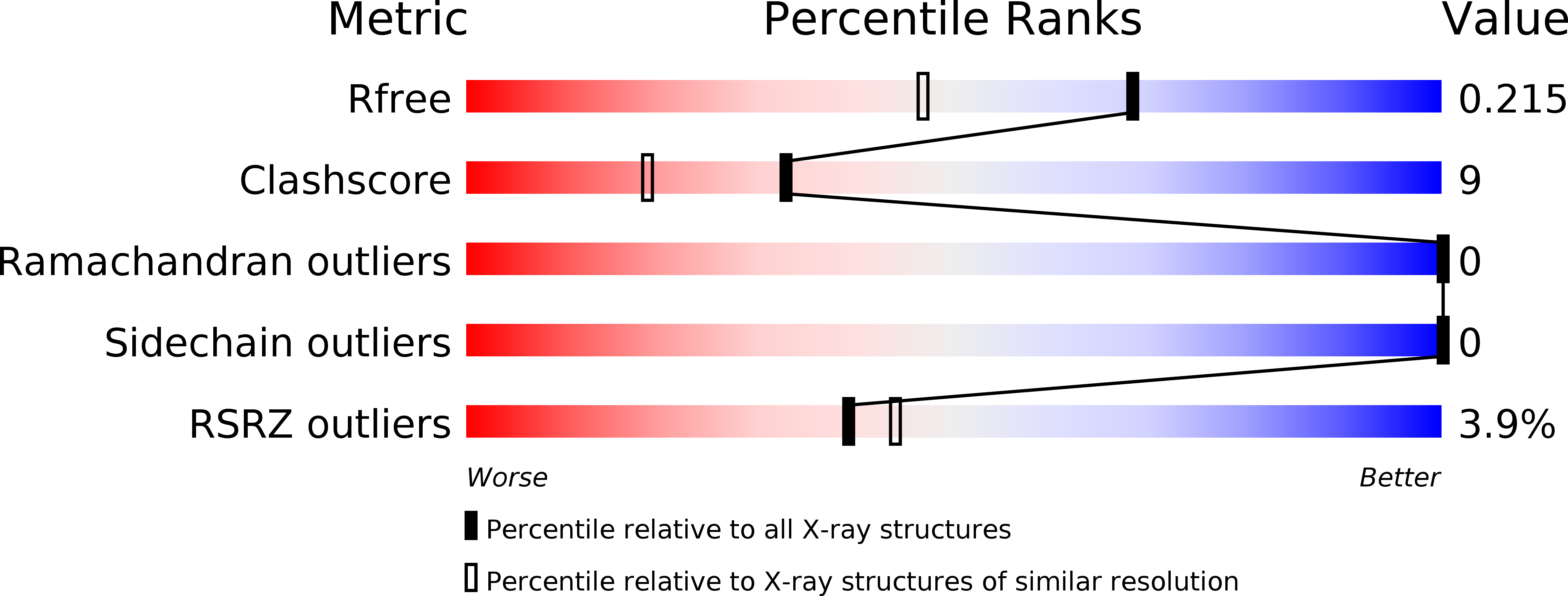
5KAZ image
Deposition Date 2016-06-02
Release Date 2017-04-12
Last Version Date 2024-02-28
Entry Detail
PDB ID:
5KAZ
Title:
Human SH2D1B structure
Biological Source:
Source Organism:
Homo sapiens (Taxon ID: 9606)
Host Organism:
Method Details:
Experimental Method:
Resolution:
1.70 Å
R-Value Free:
0.21
R-Value Work:
0.18
R-Value Observed:
0.19
Space Group:
I 4 2 2
Macromolecular Entities
Polymer Type:polypeptide(L)
Molecule:SH2 domain-containing protein 1B
Gene (Uniprot):SH2D1B
Chain IDs:A
Chain Length:104
Number of Molecules:1
Biological Source:Homo sapiens
Ligand Molecules
Primary Citation
The X-ray Crystallographic Structure of Human EAT2 (SH2D1B).
Protein Pept. Lett. 23 862 866 (2016)
PMID: 27586300

Abstact

Ewing's Sarcoma transcript-2 (EAT2) also known as SH2D1B is involved in regulation of signalling lymphocytic activation molecule (SLAM) family receptor functions. Cytoplasmic tails of SLAM family receptors contain tyrosine residues which mediate the downstream signal transduction through their phosphorylation. EAT2, composed of a single SH2 domain and a short C-terminal tail, binds to the phosphotyrosine residues and regulates SLAM family receptor signalling. We have determined the crystal structure of the human EAT2 protein in an unliganded form. Compared with the mouse EAT2-peptide complex structure, we observe conformational differences in the loops involved in ligand binding. When compared with SAP, the other single SH2 domain protein in human, EAT2 shows similar binding energies to unphosphorylated ligands. This is inconsistent to the previous data showing low affinity of EAT2 toward unphosphorylated peptides compared to SAP which shows high affinity. Additional factors other than the SH2 domains may contribute to the reported differences.

Legend

Protein

Chemical

Disease

Primary Citation of related structures