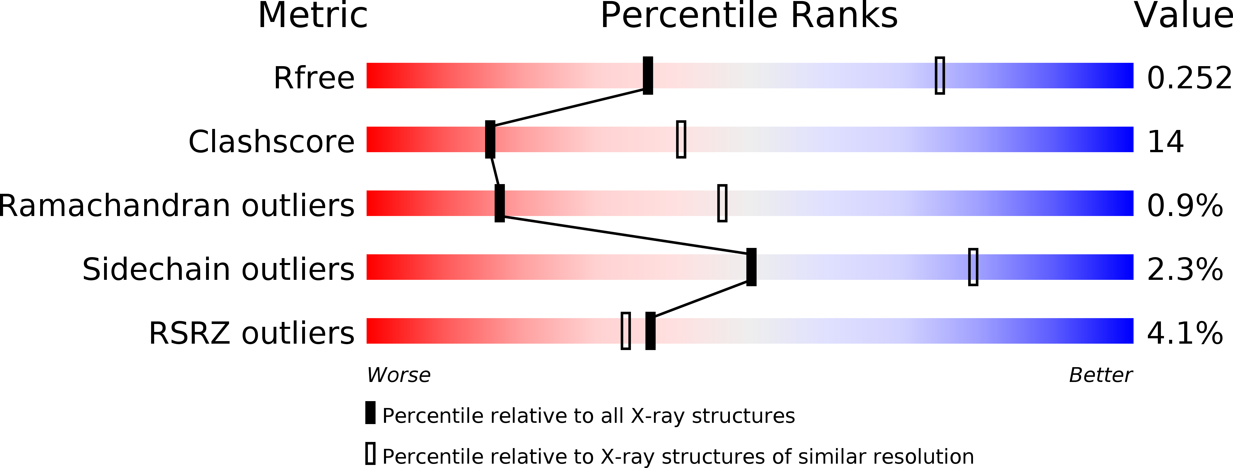
5K82 image
Deposition Date 2016-05-27
Release Date 2016-08-10
Last Version Date 2023-09-27
Entry Detail
PDB ID:
5K82
Keywords:
Title:
Crystal Structure of a Primate APOBEC3G N-Terminal Domain
Biological Source:
Source Organism:
Macaca mulatta (Taxon ID: 9544)
Host Organism:
Method Details:
Experimental Method:
Resolution:
2.91 Å
R-Value Free:
0.25
R-Value Work:
0.22
R-Value Observed:
0.22
Space Group:
P 32
Macromolecular Entities
Polymer Type:polypeptide(L)
Molecule:Apolipoprotein B mRNA editing enzyme, catalytic peptide-like 3G,Apolipoprotein B mRNA editing enzyme, catalytic peptide-like 3G
Gene (Uniprot):APOBEC3G
Chain IDs:A, B, C, D
Chain Length:196
Number of Molecules:4
Biological Source:Macaca mulatta
Ligand Molecules
Primary Citation
Crystal structures of APOBEC3G N-domain alone and its complex with DNA.
Nat Commun 7 12193 12193 (2016)
PMID: 27480941 DOI: 10.1038/ncomms12193

Abstact

APOBEC3G (A3G) is a potent restriction factor of HIV-1. The N-terminal domain of A3G (A3G-CD1) is responsible for oligomerization and nucleic acid binding, both of which are essential for anti-HIV activity. As a countermeasure, HIV-1 viral infectivity factor (Vif) binds A3G-CD1 to mediate A3G degradation. The structural basis for the functions of A3G-CD1 remains elusive. Here, we report the crystal structures of a primate A3G-CD1 (rA3G-CD1) alone and in complex with single-stranded DNA (ssDNA). rA3G-CD1 shares a conserved core structure with the previously determined catalytic APOBECs, but displays unique features for surface charge, dimerization and nucleic acid binding. Its co-crystal structure with ssDNA reveals how the conformations of loops and residues surrounding the Zn-coordinated centre (Zn-centre) change upon DNA binding. The dimerization interface of rA3G-CD1 is important for oligomerization, nucleic acid binding and Vif-mediated degradation. These findings elucidate the molecular basis of antiviral mechanism and HIV-Vif targeting of A3G.

Legend

Protein

Chemical

Disease

Primary Citation of related structures