5K4Y image
Entry Detail
PDB ID:
5K4Y
Keywords:
Title:
Three-dimensional structure of L-threonine 3-dehydrogenase from Trypanosoma brucei refined to 1.77 angstroms
Biological Source:
Source Organism:
Host Organism:
PDB Version:
Deposition Date:
2016-05-22
Release Date:
2018-01-17
Method Details:
Experimental Method:
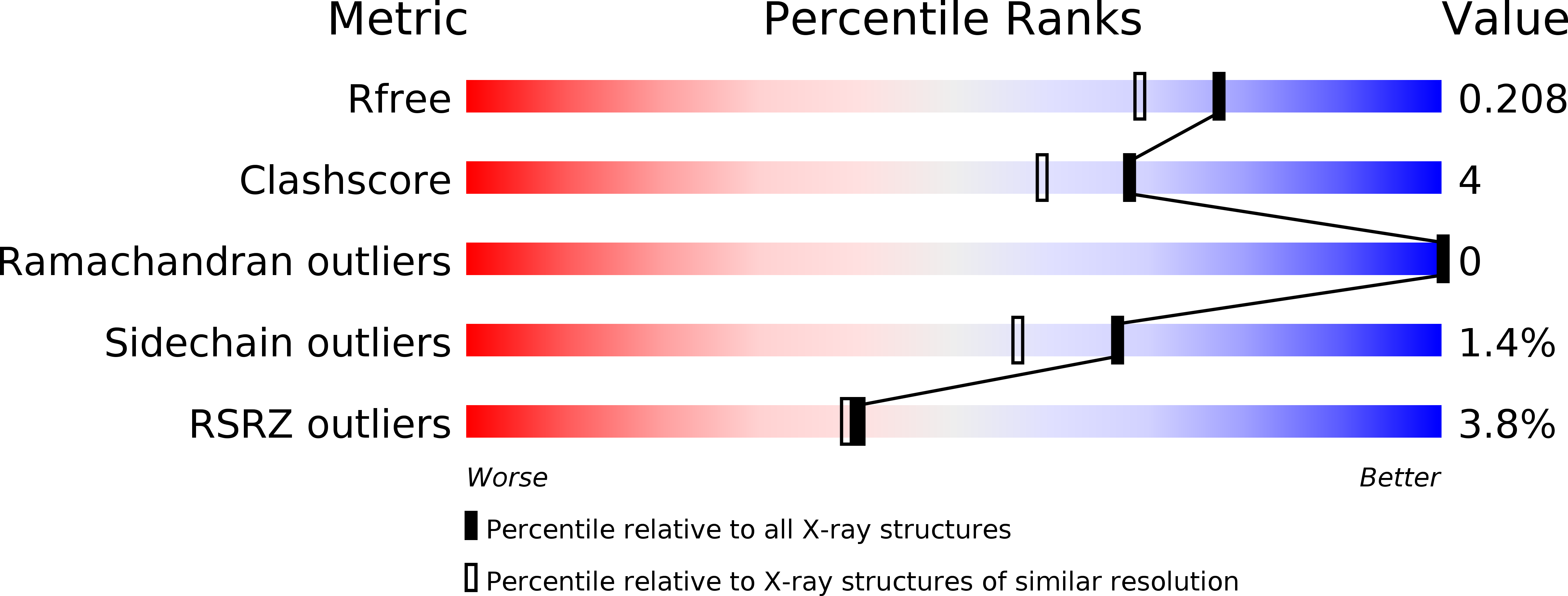
Resolution:
1.77 Å
R-Value Free:
0.20
R-Value Work:
0.16
R-Value Observed:
0.16
Space Group:
P 21 21 2
Macromolecular Entities
Polymer Type:polypeptide(L)
Description:L-threonine 3-dehydrogenase
Chain IDs:A, B, C, D, E, F
Chain Length:320
Number of Molecules:6
Biological Source:Trypanosoma brucei
Primary Citation
Structure and function of L-threonine-3-dehydrogenase from the parasitic protozoan Trypanosoma brucei revealed by X-ray crystallography and geometric simulations.
Acta Crystallogr D Struct Biol 74 861 876 (2018)
PMID: 30198897 DOI: 10.1107/S2059798318009208

Abstact

Two of the world's most neglected tropical diseases, human African trypanosomiasis (HAT) and Chagas disease, are caused by protozoan parasites of the genus Trypanosoma. These organisms possess specialized metabolic pathways, frequently distinct from those in humans, which have potential to be exploited as novel drug targets. This study elucidates the structure and function of L-threonine-3-dehydrogenase (TDH) from T. brucei, the causative pathogen of HAT. TDH is a key enzyme in the metabolism of L-threonine, and an inhibitor of TDH has been shown to have trypanocidal activity in the procyclic form of T. brucei. TDH is a nonfunctional pseudogene in humans, suggesting that it may be possible to rationally design safe and specific therapies for trypanosomiasis by targeting this parasite enzyme. As an initial step, the TDH gene from T. brucei was expressed and the three-dimensional structure of the enzyme was solved by X-ray crystallography. In multiple crystallographic structures, T. brucei TDH is revealed to be a dimeric short-chain dehydrogenase that displays a considerable degree of conformational variation in its ligand-binding regions. Geometric simulations of the structure have provided insight into the dynamic behaviour of this enzyme. Furthermore, structures of TDH bound to its natural substrates and known inhibitors have been determined, giving an indication of the mechanism of catalysis of the enzyme. Collectively, these results provide vital details for future drug design to target TDH or related enzymes.

Legend

Protein

Chemical

Disease

Primary Citation of related structures