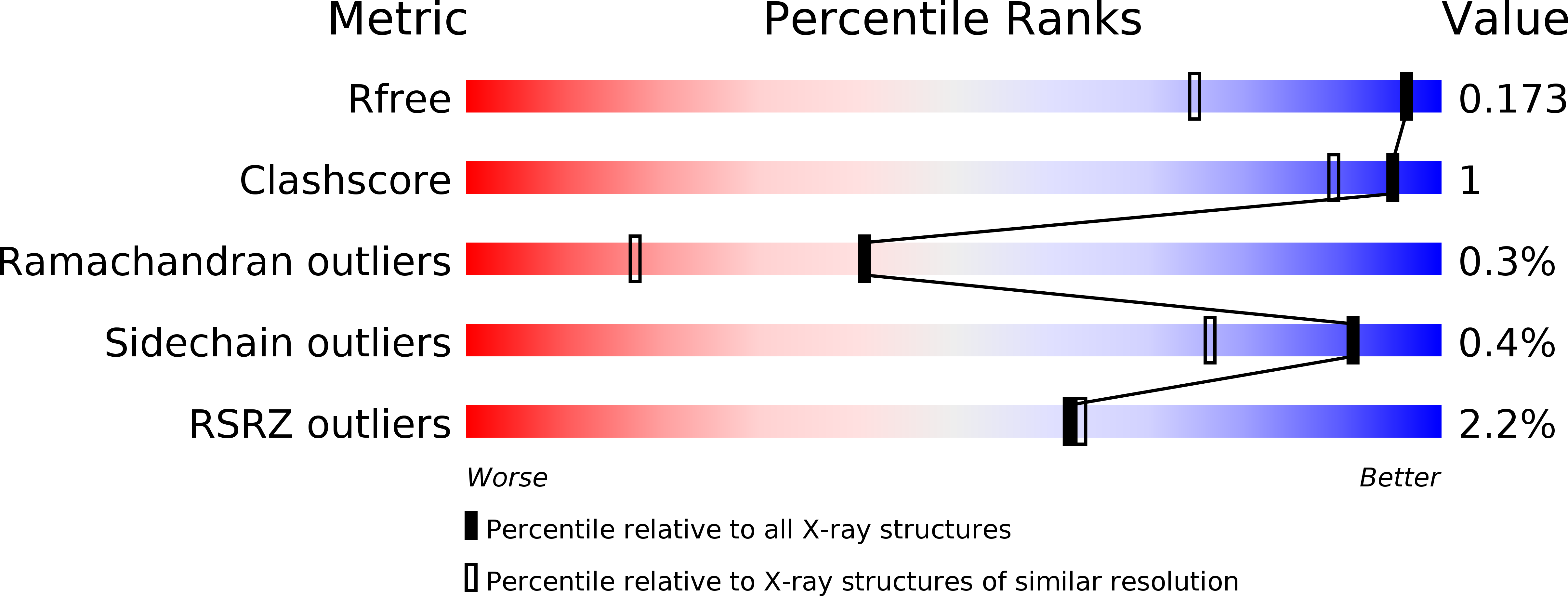
5K4P image
Deposition Date 2016-05-21
Release Date 2016-08-31
Last Version Date 2024-10-09
Entry Detail
PDB ID:
5K4P
Keywords:
Title:
Catalytic Domain of MCR-1 phosphoethanolamine transferase
Biological Source:
Source Organism:
Method Details:
Experimental Method:
Resolution:
1.32 Å
R-Value Free:
0.17
R-Value Work:
0.15
R-Value Observed:
0.15
Space Group:
P 43 21 2
Macromolecular Entities
Structures with similar UniProt ID
Protein Blast
Polymer Type:polypeptide(L)
Molecule:Probable phosphatidylethanolamine transferase Mcr-1
Gene (Uniprot):mcr1
Chain IDs:A
Chain Length:342
Number of Molecules:1
Biological Source:Escherichia coli BL21(DE3)
Modified Residue
Compound ID Chain ID Parent Comp ID Details 2D Image
TPO A THR modified residue
Primary Citation
Structure of the catalytic domain of the colistin resistance enzyme MCR-1.
Bmc Biol. 14 81 81 (2016)
PMID: 27655155 DOI: 10.1186/s12915-016-0303-0

Abstact

BACKGROUND Due to the paucity of novel antibiotics, colistin has become a last resort antibiotic for treating multidrug resistant bacteria. Colistin acts by binding the lipid A component of lipopolysaccharides and subsequently disrupting the bacterial membrane. The recently identified plasmid-encoded MCR-1 enzyme is the first transmissible colistin resistance determinant and is a cause for concern for the spread of this resistance trait. MCR-1 is a phosphoethanolamine transferase that catalyzes the addition of phosphoethanolamine to lipid A to decrease colistin affinity. RESULTS The structure of the catalytic domain of MCR-1 at 1.32 Å reveals the active site is similar to that of related phosphoethanolamine transferases. CONCLUSIONS The putative nucleophile for catalysis, threonine 285, is phosphorylated in cMCR-1 and a zinc is present at a conserved site in addition to three zincs more peripherally located in the active site. As noted for catalytic domains of other phosphoethanolamine transferases, binding sites for the lipid A and phosphatidylethanolamine substrates are not apparent in the cMCR-1 structure, suggesting that they are present in the membrane domain.

Legend

Protein

Chemical

Disease

Primary Citation of related structures