5K3H image
Deposition Date 2016-05-19
Release Date 2016-08-24
Last Version Date 2023-09-27
Entry Detail
PDB ID:
5K3H
Keywords:
Title:
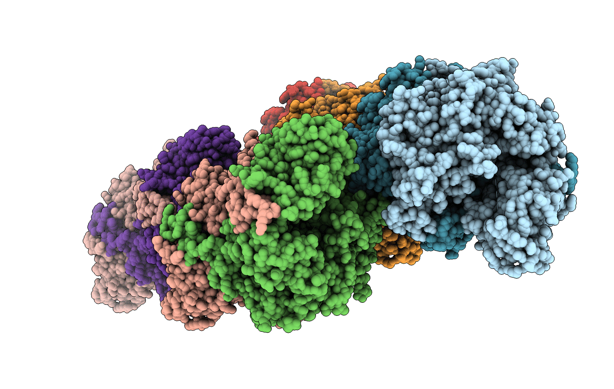
Crystals structure of Acyl-CoA oxidase-1 in Caenorhabditis elegans, Apo form-II
Biological Source:
Source Organism:
Host Organism:
Method Details:
Experimental Method:
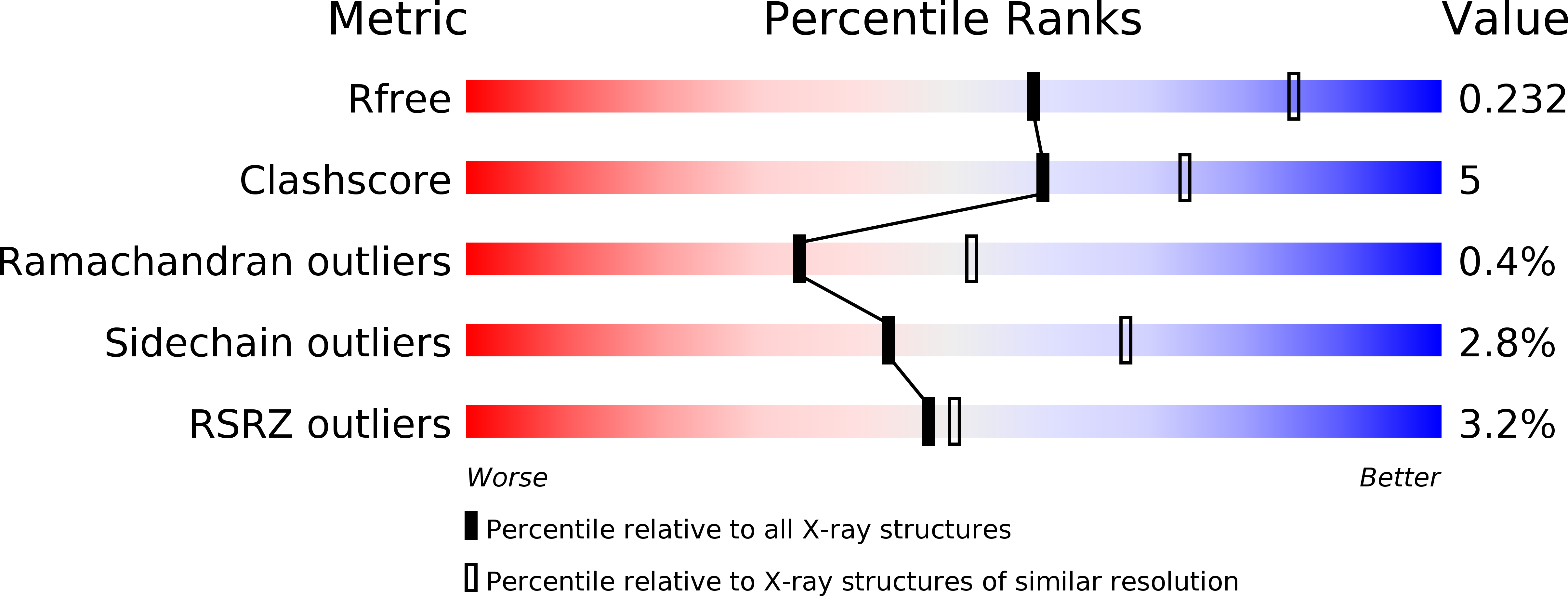
Resolution:
2.48 Å
R-Value Free:
0.23
R-Value Work:
0.19
R-Value Observed:
0.19
Space Group:
P 1 21 1
Macromolecular Entities
Structures with similar UniProt ID
Protein Blast
Polymer Type:polypeptide(L)
Molecule:Acyl-coenzyme A oxidase
Gene (Uniprot):acox-1.1
Chain IDs:A, B, C, D, E, F, G, H
Chain Length:684
Number of Molecules:8
Biological Source:Caenorhabditis elegans
Primary Citation
Structural characterization of acyl-CoA oxidases reveals a direct link between pheromone biosynthesis and metabolic state in Caenorhabditis elegans.
Proc.Natl.Acad.Sci.USA 113 10055 10060 (2016)
PMID: 27551084 DOI: 10.1073/pnas.1608262113

Abstact

Caenorhabditis elegans secretes ascarosides as pheromones to communicate with other worms and to coordinate the development and behavior of the population. Peroxisomal β-oxidation cycles shorten the side chains of ascaroside precursors to produce the short-chain ascaroside pheromones. Acyl-CoA oxidases, which catalyze the first step in these β-oxidation cycles, have different side chain-length specificities and enable C. elegans to regulate the production of specific ascaroside pheromones. Here, we determine the crystal structure of the acyl-CoA oxidase 1 (ACOX-1) homodimer and the ACOX-2 homodimer bound to its substrate. Our results provide a molecular basis for the substrate specificities of the acyl-CoA oxidases and reveal why some of these enzymes have a very broad substrate range, whereas others are quite specific. Our results also enable predictions to be made for the roles of uncharacterized acyl-CoA oxidases in C. elegans and in other nematode species. Remarkably, we show that most of the C. elegans acyl-CoA oxidases that participate in ascaroside biosynthesis contain a conserved ATP-binding pocket that lies at the dimer interface, and we identify key residues in this binding pocket. ATP binding induces a structural change that is associated with tighter binding of the FAD cofactor. Mutations that disrupt ATP binding reduce FAD binding and reduce enzyme activity. Thus, ATP may serve as a regulator of acyl-CoA oxidase activity, thereby directly linking ascaroside biosynthesis to ATP concentration and metabolic state.

Legend

Protein

Chemical

Disease

Primary Citation of related structures
Feedback Form
Name
Email
Institute
Feedback