Deposition Date
2016-05-19
Release Date
2017-04-12
Last Version Date
2024-10-09
Entry Detail
PDB ID:
5K33
Keywords:
Title:
Crystal structure of extracellular domain of HER2 in complex with Fcab STAB19
Biological Source:
Source Organism(s):
Homo sapiens (Taxon ID: 9606)
Expression System(s):
Method Details:
Experimental Method:
Resolution:
3.30 Å
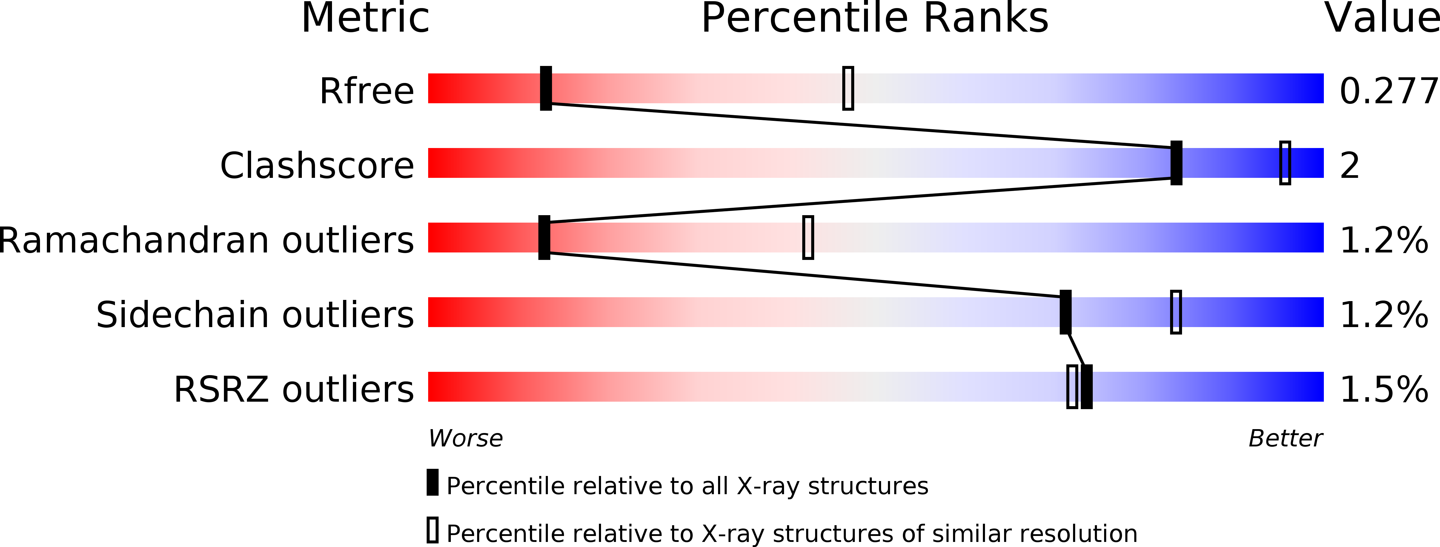
R-Value Free:
0.27
R-Value Work:
0.21
R-Value Observed:
0.22
Space Group:
P 32 2 1