5K26 image
Entry Detail
PDB ID:
5K26
Keywords:
Title:
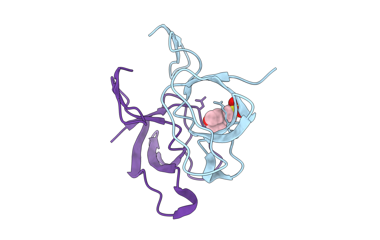
Structure of the SH3 domain of MLK3 bound to peptide generated from phage display
Biological Source:
Source Organism:
Host Organism:
PDB Version:
Deposition Date:
2016-05-18
Release Date:
2017-12-13
Method Details:
Experimental Method:
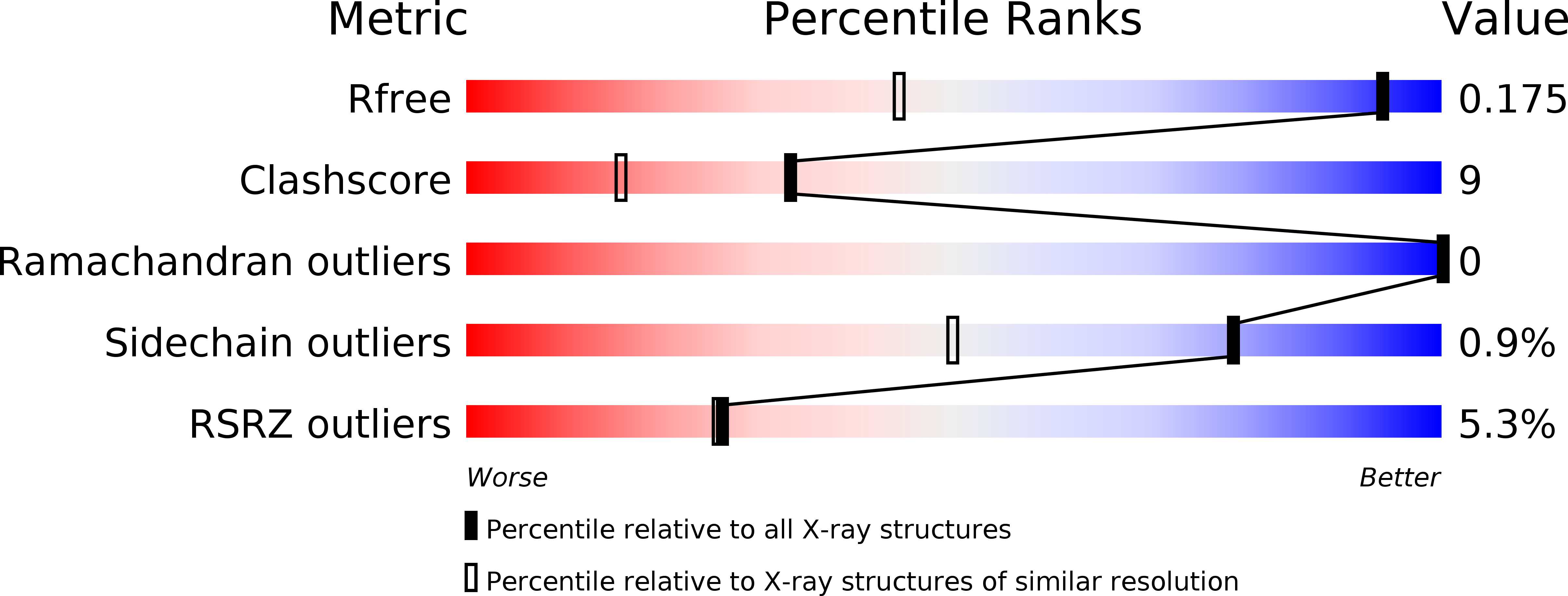
Resolution:
1.20 Å
R-Value Free:
0.16
R-Value Work:
0.13
R-Value Observed:
0.13
Space Group:
P 31
Macromolecular Entities
Polymer Type:polypeptide(L)
Description:Mitogen-activated protein kinase kinase kinase 11,Chimera protein of MLK3-SH3 and MIP
Chain IDs:A, B
Chain Length:86
Number of Molecules:2
Biological Source:Homo sapiens
Ligand Molecules
Primary Citation
Identification of two distinct peptide-binding pockets in the SH3 domain of human mixed-lineage kinase 3.
J. Biol. Chem. 293 13553 13565 (2018)
PMID: 29980598 DOI: 10.1074/jbc.RA117.000262

Abstact

Mixed-lineage kinase 3 (MLK3; also known as MAP3K11) is a Ser/Thr protein kinase widely expressed in normal and cancerous tissues, including brain, lung, liver, heart, and skeletal muscle tissues. Its Src homology 3 (SH3) domain has been implicated in MLK3 autoinhibition and interactions with other proteins, including those from viruses. The MLK3 SH3 domain contains a six-amino-acid insert corresponding to the n-Src insert, suggesting that MLK3 may bind additional peptides. Here, affinity selection of a phage-displayed combinatorial peptide library for MLK3's SH3 domain yielded a 13-mer peptide, designated "MLK3 SH3-interacting peptide" (MIP). Unlike most SH3 domain peptide ligands, MIP contained a single proline. The 1.2-Å crystal structure of the MIP-bound SH3 domain revealed that the peptide adopts a β-hairpin shape, and comparison with a 1.5-Å apo SH3 domain structure disclosed that the n-Src loop in SH3 undergoes an MIP-induced conformational change. A 1.5-Å structure of the MLK3 SH3 domain bound to a canonical proline-rich peptide from hepatitis C virus nonstructural 5A (NS5A) protein revealed that it and MIP bind the SH3 domain at two distinct sites, but biophysical analyses suggested that the two peptides compete with each other for SH3 binding. Moreover, SH3 domains of MLK1 and MLK4, but not MLK2, also bound MIP, suggesting that the MLK1-4 family may be differentially regulated through their SH3 domains. In summary, we have identified two distinct peptide-binding sites in the SH3 domain of MLK3, providing critical insights into mechanisms of ligand binding by the MLK family of kinases.

Legend

Protein

Chemical

Disease

Primary Citation of related structures