Deposition Date
2016-05-18
Release Date
2016-11-16
Last Version Date
2024-03-06
Entry Detail
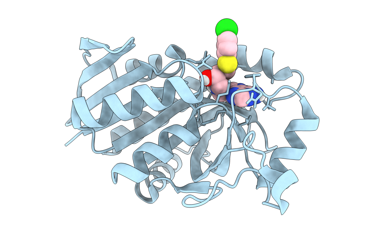
PDB ID:
5K1Z
Keywords:
Title:
Joint X-ray/neutron structure of MTAN complex with p-ClPh-Thio-DADMe-ImmA
Biological Source:
Source Organism(s):
Helicobacter pylori (Taxon ID: 210)
Expression System(s):
Method Details:
Experimental Method:
R-Value Free:
['0.28
R-Value Work:
['0.25
R-Value Observed:
['?', '?'].00
Space Group:
P 32 2 1