5K1V image
Deposition Date 2016-05-18
Release Date 2017-03-29
Last Version Date 2024-10-09
Entry Detail
PDB ID:
5K1V
Keywords:
Title:
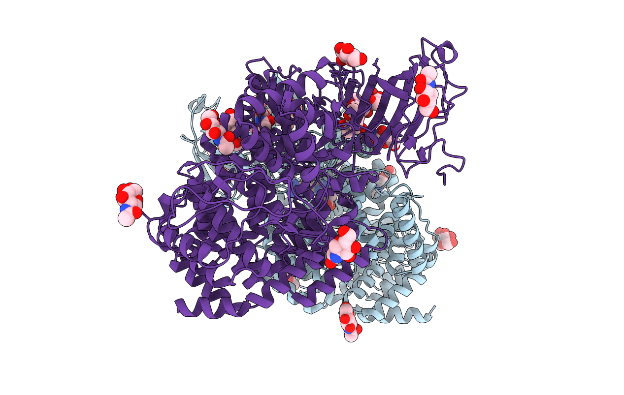
Crystal structure of Endoplasmic Reticulum aminopeptidase 2 (ERAP2) in complex with a diaminobenzoic acid derivative ligand.
Biological Source:
Source Organism:
Homo sapiens (Taxon ID: 9606)
Host Organism:
Method Details:
Experimental Method:
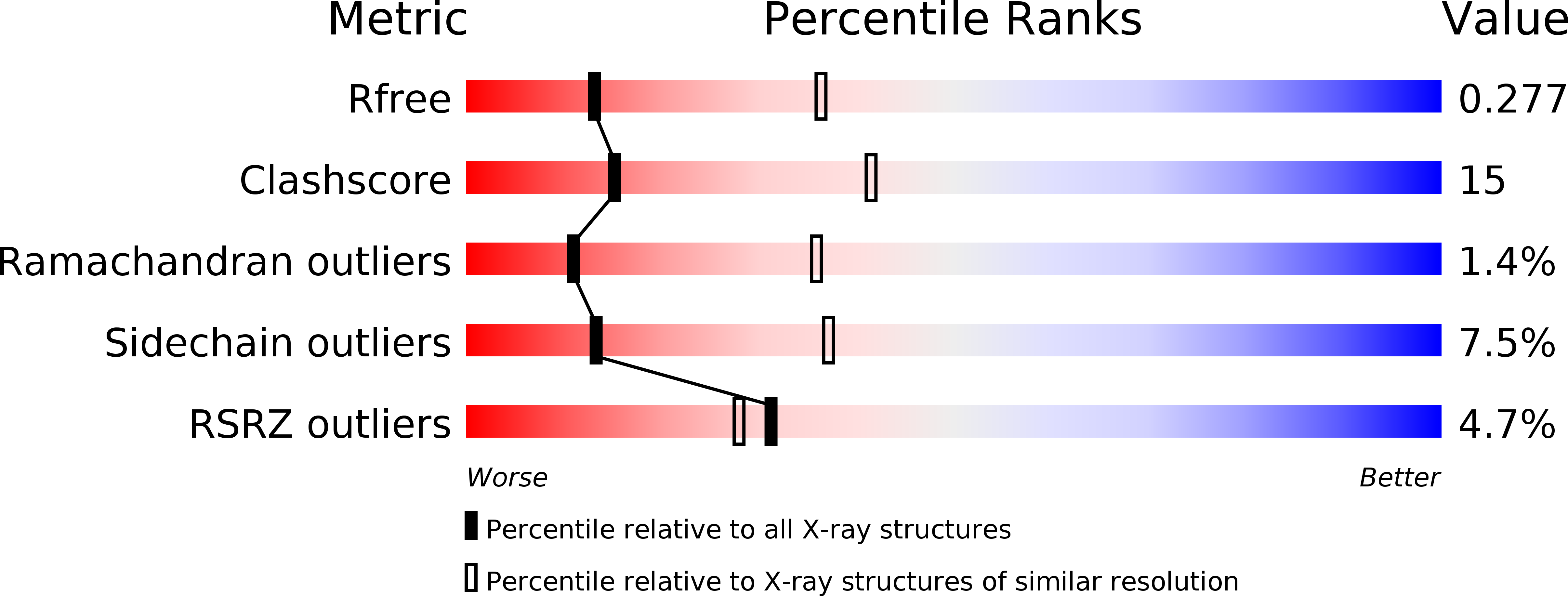
Resolution:
2.90 Å
R-Value Free:
0.27
R-Value Work:
0.20
R-Value Observed:
0.20
Space Group:
P 1 21 1
Macromolecular Entities
Polymer Type:polypeptide(L)
Molecule:Endoplasmic reticulum aminopeptidase 2
Gene (Uniprot):ERAP2
Chain IDs:A, B
Chain Length:967
Number of Molecules:2
Biological Source:Homo sapiens
Primary Citation
Crystal Structures of ERAP2 Complexed with Inhibitors Reveal Pharmacophore Requirements for Optimizing Inhibitor Potency.
ACS Med Chem Lett 8 333 337 (2017)
PMID: 28337326 DOI: 10.1021/acsmedchemlett.6b00505

Abstact

Endoplasmic reticulum aminopeptidase 2 assists with the generation of antigenic peptides for presentation onto Major Histocompatibility Class I molecules in humans. Recent evidence has suggested that the activity of ERAP2 may contribute to the generation of autoimmunity, thus making ERAP2 a possible pharmacological target for the regulation of adaptive immune responses. To better understand the structural elements of inhibitors that govern their binding affinity to the ERAP2 active site, we cocrystallized ERAP2 with a medium activity 3,4-diaminobenzoic acid inhibitor and a poorly active hydroxamic acid derivative. Comparison of these two crystal structures with a previously solved structure of ERAP2 in complex with a potent phosphinic pseudopeptide inhibitor suggests that engaging the substrate N-terminus recognition properties of the active site is crucial for inhibitor binding even in the absence of a potent zinc-binding group. Proper utilization of all five major pharmacophores is necessary, however, to optimize inhibitor potency.

Legend

Protein

Chemical

Disease

Primary Citation of related structures