5JYX image
Deposition Date 2016-05-15
Release Date 2016-11-09
Last Version Date 2024-11-06
Entry Detail
PDB ID:
5JYX
Keywords:
Title:
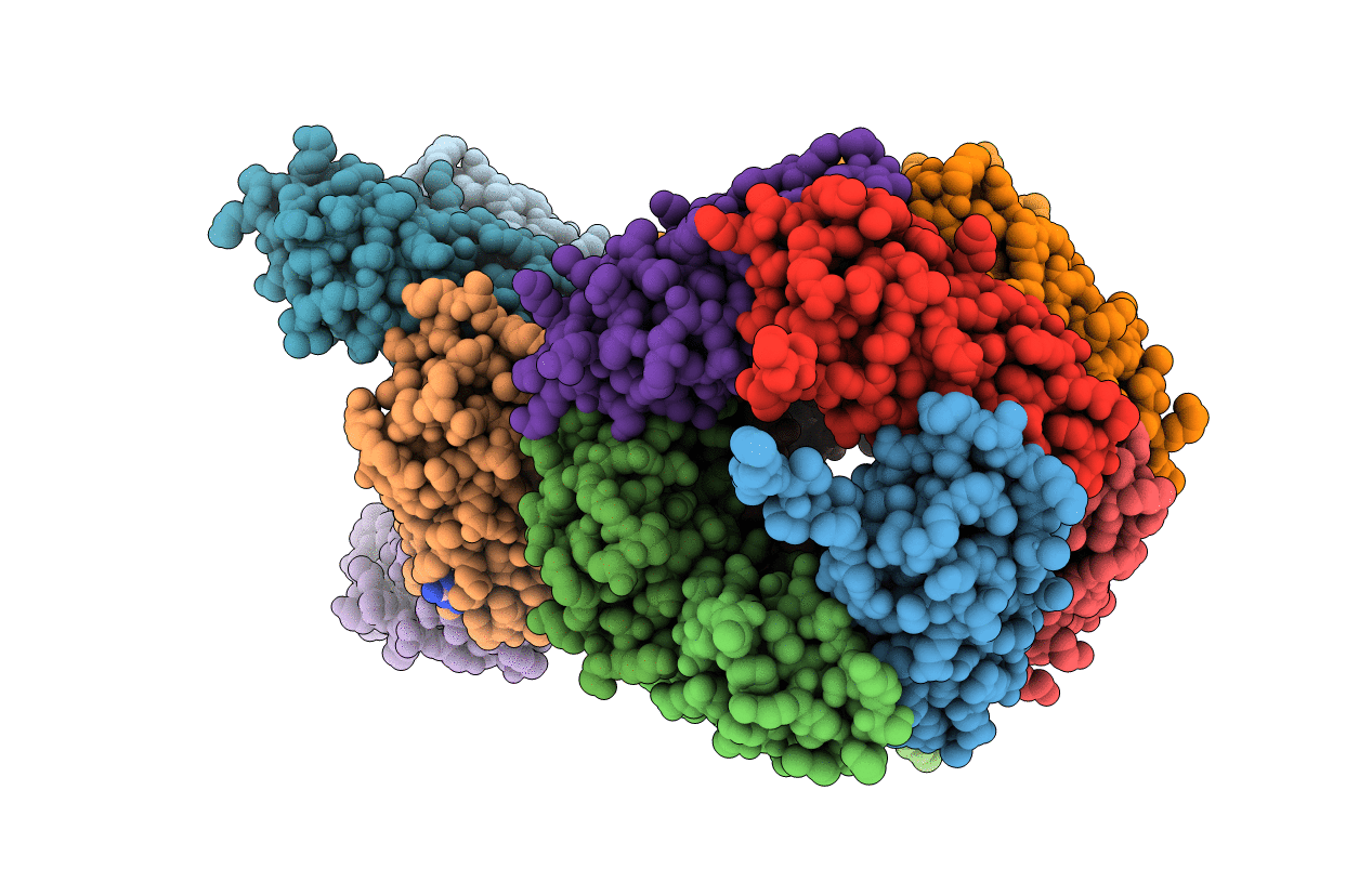
Crystal structure of the covalent thioimide intermediate of the archaeosine synthase QueF-Like
Biological Source:
Source Organism:
Host Organism:
Method Details:
Experimental Method:
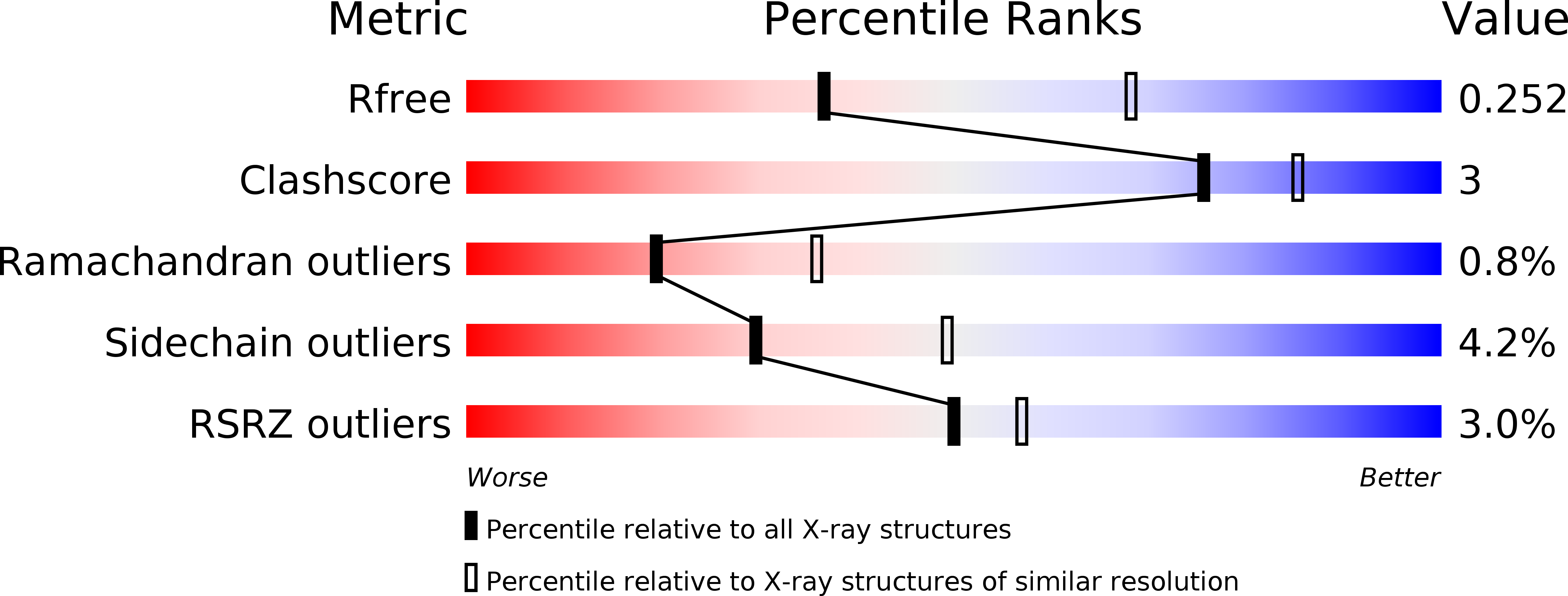
Resolution:
2.74 Å
R-Value Free:
0.25
R-Value Work:
0.17
R-Value Observed:
0.17
Space Group:
C 1 2 1
Macromolecular Entities
Polymer Type:polypeptide(L)
Molecule:Archeaosine synthase QueF-Like
Gene (Uniprot):queF-L
Chain IDs:A, B, C, D, E, F, G, H, I, J, K, L, M, N, O
Chain Length:109
Number of Molecules:15
Biological Source:Pyrobaculum calidifontis
Modified Residue
Compound ID Chain ID Parent Comp ID Details 2D Image
MSE A MET modified residue
Primary Citation
Crystal structure of the archaeosine synthase QueF-like-Insights into amidino transfer and tRNA recognition by the tunnel fold.
Proteins 85 103 116 (2017)
PMID: 27802572 DOI: 10.1002/prot.25202

Abstact

The tunneling-fold (T-fold) structural superfamily has emerged as a versatile protein scaffold of diverse catalytic activities. This is especially evident in the pathways to the 7-deazaguanosine modified nucleosides of tRNA queuosine and archaeosine. Four members of the T-fold superfamily have been confirmed in these pathways and here we report the crystal structure of a fifth enzyme; the recently discovered amidinotransferase QueF-Like (QueF-L), responsible for the final step in the biosynthesis of archaeosine in the D-loop of tRNA in a subset of Crenarchaeota. QueF-L catalyzes the conversion of the nitrile group of the 7-cyano-7-deazaguanine (preQ0) base of preQ0 -modified tRNA to a formamidino group. The structure, determined in the presence of preQ0 , reveals a symmetric T-fold homodecamer of two head-to-head facing pentameric subunits, with 10 active sites at the inter-monomer interfaces. Bound preQ0 forms a stable covalent thioimide bond with a conserved active site cysteine similar to the intermediate previously observed in the nitrile reductase QueF. Despite distinct catalytic functions, phylogenetic distributions, and only 19% sequence identity, the two enzymes share a common preQ0 binding pocket, and likely a common mechanism of thioimide formation. However, due to tight twisting of its decamer, QueF-L lacks the NADPH binding site present in QueF. A large positively charged molecular surface and a docking model suggest simultaneous binding of multiple tRNA molecules and structure-specific recognition of the D-loop by a surface groove. The structure sheds light on the mechanism of nitrile amidation, and the evolution of diverse chemistries in a common fold. Proteins 2016; 85:103-116. © 2016 Wiley Periodicals, Inc.

Legend

Protein

Chemical

Disease

Primary Citation of related structures