
Deposition Date
2016-05-13
Release Date
2017-05-24
Last Version Date
2024-01-10
Entry Detail
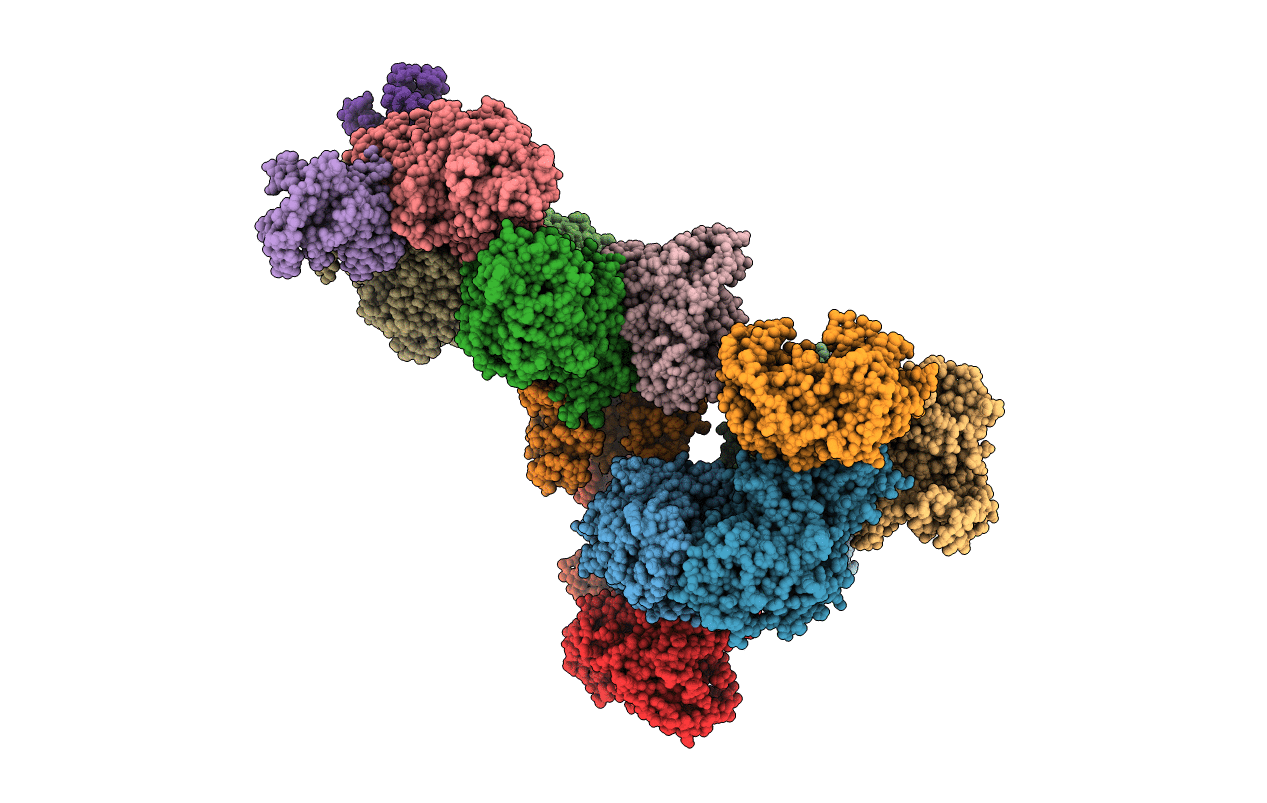
PDB ID:
5JY7
Keywords:
Title:
Complex of Mycobacterium smegmatis trehalose synthase with maltokinase
Biological Source:
Source Organism(s):
Expression System(s):
Method Details:
Experimental Method:
Resolution:
3.60 Å
R-Value Free:
0.28
R-Value Work:
0.26
R-Value Observed:
0.26
Space Group:
P 43
