5JWO image
Deposition Date 2016-05-12
Release Date 2017-03-29
Last Version Date 2024-03-06
Entry Detail
PDB ID:
5JWO
Title:
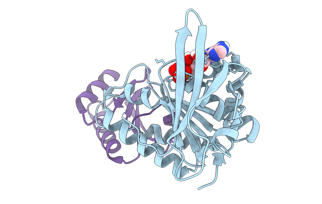
Crystal structure of foldswitch-stabilized KaiB in complex with the N-terminal CI domain of KaiC from Thermosynechococcus elongatus
Biological Source:
Source Organism:
Method Details:
Experimental Method:
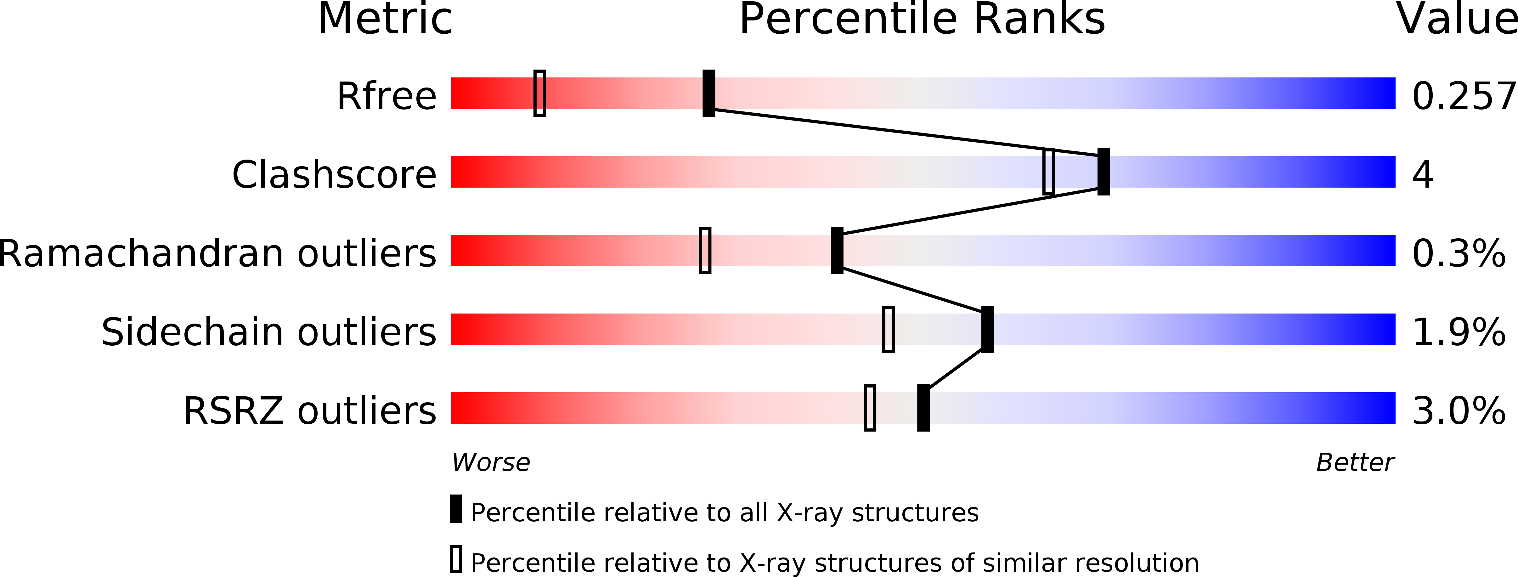
Resolution:
1.80 Å
R-Value Free:
0.25
R-Value Work:
0.21
R-Value Observed:
0.22
Space Group:
P 2 21 21
Macromolecular Entities
Polymer Type:polypeptide(L)
Molecule:Circadian clock protein kinase KaiC
Gene (Uniprot):kaiC
Mutations:R41A, K173A
Chain IDs:A
Chain Length:247
Number of Molecules:1
Biological Source:Thermosynechococcus elongatus
Polymer Type:polypeptide(L)
Molecule:Circadian clock protein KaiB
Gene (Uniprot):kaiB
Mutations:Y8A, G89A, D91R, Y94A
Chain IDs:B
Chain Length:99
Number of Molecules:1
Biological Source:Thermosynechococcus elongatus (strain BP-1)
Ligand Molecules
Primary Citation
Structural basis of the day-night transition in a bacterial circadian clock.
Science 355 1174 1180 (2017)
PMID: 28302851 DOI: 10.1126/science.aag2516

Abstact

Circadian clocks are ubiquitous timing systems that induce rhythms of biological activities in synchrony with night and day. In cyanobacteria, timing is generated by a posttranslational clock consisting of KaiA, KaiB, and KaiC proteins and a set of output signaling proteins, SasA and CikA, which transduce this rhythm to control gene expression. Here, we describe crystal and nuclear magnetic resonance structures of KaiB-KaiC,KaiA-KaiB-KaiC, and CikA-KaiB complexes. They reveal how the metamorphic properties of KaiB, a protein that adopts two distinct folds, and the post-adenosine triphosphate hydrolysis state of KaiC create a hub around which nighttime signaling events revolve, including inactivation of KaiA and reciprocal regulation of the mutually antagonistic signaling proteins, SasA and CikA.

Legend

Protein

Chemical

Disease

Primary Citation of related structures