5JVO image
Deposition Date 2016-05-11
Release Date 2016-08-31
Last Version Date 2023-09-27
Entry Detail
PDB ID:
5JVO
Title:
Crystal structure of the Arginine Repressor from the pathogenic bacterium Corynebacterium pseudotuberculosis
Biological Source:
Source Organism:
Host Organism:
Method Details:
Experimental Method:
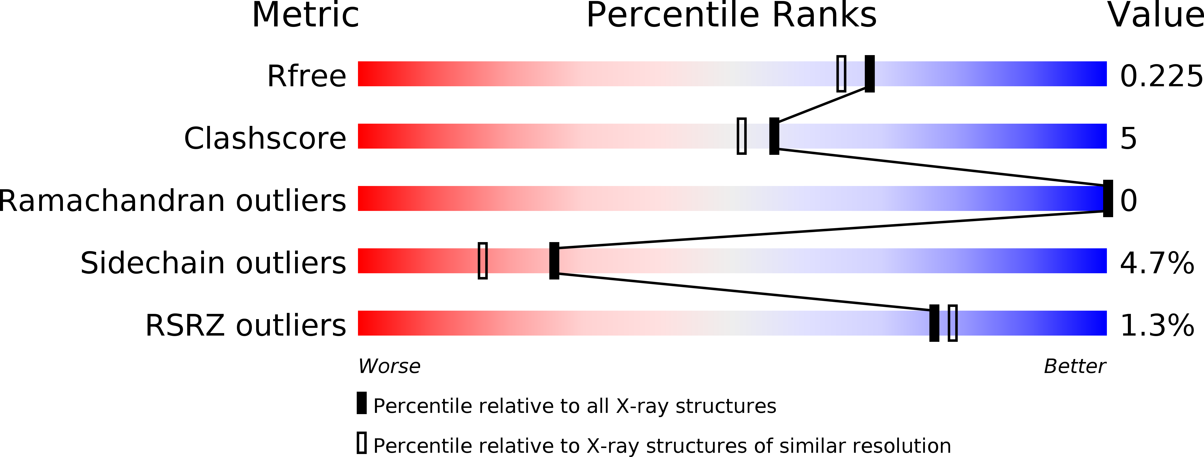
Resolution:
1.90 Å
R-Value Free:
0.22
R-Value Work:
0.17
R-Value Observed:
0.18
Space Group:
P 21 3
Macromolecular Entities
Structures with similar UniProt ID
Protein Blast
Polymer Type:polypeptide(L)
Molecule:Arginine repressor
Chain IDs:A, B
Chain Length:97
Number of Molecules:2
Biological Source:Corynebacterium pseudotuberculosis
Primary Citation
Tyrosine binding and promiscuity in the arginine repressor from the pathogenic bacterium Corynebacterium pseudotuberculosis.
Biochem.Biophys.Res.Commun. 475 350 355 (2016)
PMID: 27233609 DOI: 10.1016/j.bbrc.2016.05.091

Abstact

The arginine repressor (ArgR) regulates arginine biosynthesis in a number of microorganisms and consists of two domains interlinked by a short peptide; the N-terminal domain is involved in DNA binding and the C-terminal domain binds arginine and forms a hexamer made-up of a dimer of trimers. The crystal structure of the C-terminal domain of ArgR from the pathogenic Corynebacterium pseudotuberculosis determined at 1.9 Å resolution contains a tightly bound tyrosine at the arginine-binding site indicating hitherto unobserved promiscuity. Structural analysis of the binding pocket displays clear molecular adaptations to accommodate tyrosine binding suggesting the possible existence of an alternative regulatory process in this pathogenic bacterium.

Legend

Protein

Chemical

Disease

Primary Citation of related structures