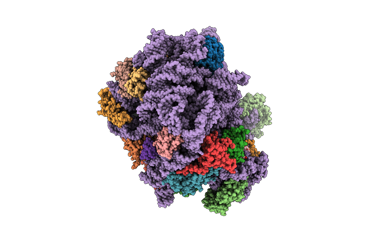
5JVH image
Entry Detail
PDB ID:
5JVH
Keywords:
Title:
The crystal structure large ribosomal subunit (50S) of Deinococcus radiodurans in complex with evernimicin
Biological Source:
PDB Version:
Deposition Date:
2016-05-11
Release Date:
2016-11-09
Method Details:
Experimental Method:
Resolution:
3.58 Å
R-Value Free:
0.24
R-Value Work:
0.20
R-Value Observed:
0.20
Space Group:
I 2 2 2
Macromolecular Entities
Polymer Type:polypeptide(L)
Description:50S ribosomal protein L33
Chain IDs:Z (auth: 1)
Chain Length:54
Number of Molecules:1
Biological Source:Deinococcus radiodurans R1
Polymer Type:polypeptide(L)
Description:50S ribosomal protein L34
Chain IDs:AA (auth: 2)
Chain Length:47
Number of Molecules:1
Biological Source:Deinococcus radiodurans R1
Polymer Type:polypeptide(L)
Description:50S ribosomal protein L35
Chain IDs:BA (auth: 3)
Chain Length:66
Number of Molecules:1
Biological Source:Deinococcus radiodurans R1
Polymer Type:polypeptide(L)
Description:50S ribosomal protein L2
Chain IDs:C (auth: A)
Chain Length:275
Number of Molecules:1
Biological Source:Deinococcus radiodurans R1
Polymer Type:polypeptide(L)
Description:50S ribosomal protein L3
Chain IDs:D (auth: B)
Chain Length:211
Number of Molecules:1
Biological Source:Deinococcus radiodurans R1
Polymer Type:polypeptide(L)
Description:50S ribosomal protein L4
Chain IDs:E (auth: C)
Chain Length:205
Number of Molecules:1
Biological Source:Deinococcus radiodurans R1
Polymer Type:polypeptide(L)
Description:50S ribosomal protein L5
Chain IDs:F (auth: D)
Chain Length:180
Number of Molecules:1
Biological Source:Deinococcus radiodurans R1
Polymer Type:polypeptide(L)
Description:50S ribosomal protein L6
Chain IDs:G (auth: E)
Chain Length:185
Number of Molecules:1
Biological Source:Deinococcus radiodurans R1
Polymer Type:polypeptide(L)
Description:50S ribosomal protein L13
Chain IDs:H (auth: G)
Chain Length:174
Number of Molecules:1
Biological Source:Deinococcus radiodurans R1
Polymer Type:polypeptide(L)
Description:50S ribosomal protein L14
Chain IDs:I (auth: H)
Chain Length:134
Number of Molecules:1
Biological Source:Deinococcus radiodurans R1
Polymer Type:polypeptide(L)
Description:50S ribosomal protein L15
Chain IDs:J (auth: I)
Chain Length:156
Number of Molecules:1
Biological Source:Deinococcus radiodurans R1
Polymer Type:polypeptide(L)
Description:50S ribosomal protein L16
Chain IDs:K (auth: J)
Chain Length:141
Number of Molecules:1
Biological Source:Deinococcus radiodurans R1
Polymer Type:polypeptide(L)
Description:50S ribosomal protein L17
Chain IDs:L (auth: K)
Chain Length:116
Number of Molecules:1
Biological Source:Deinococcus radiodurans R1
Polymer Type:polypeptide(L)
Description:50S ribosomal protein L18
Chain IDs:M (auth: L)
Chain Length:114
Number of Molecules:1
Biological Source:Deinococcus radiodurans R1
Polymer Type:polypeptide(L)
Description:50S ribosomal protein L19
Chain IDs:N (auth: M)
Chain Length:166
Number of Molecules:1
Biological Source:Deinococcus radiodurans R1
Polymer Type:polypeptide(L)
Description:50S ribosomal protein L20
Chain IDs:O (auth: N)
Chain Length:118
Number of Molecules:1
Biological Source:Deinococcus radiodurans R1
Polymer Type:polypeptide(L)
Description:50S ribosomal protein L21
Chain IDs:P (auth: O)
Chain Length:100
Number of Molecules:1
Biological Source:Deinococcus radiodurans R1
Polymer Type:polypeptide(L)
Description:50S ribosomal protein L22
Chain IDs:Q (auth: P)
Chain Length:134
Number of Molecules:1
Biological Source:Deinococcus radiodurans R1
Polymer Type:polypeptide(L)
Description:50S ribosomal protein L23
Chain IDs:R (auth: Q)
Chain Length:95
Number of Molecules:1
Biological Source:Deinococcus radiodurans R1
Polymer Type:polypeptide(L)
Description:50S ribosomal protein L24
Chain IDs:S (auth: R)
Chain Length:115
Number of Molecules:1
Biological Source:Deinococcus radiodurans R1
Polymer Type:polypeptide(L)
Description:50S ribosomal protein L25
Chain IDs:T (auth: S)
Chain Length:237
Number of Molecules:1
Biological Source:Deinococcus radiodurans R1
Polymer Type:polypeptide(L)
Description:50S ribosomal protein L27
Chain IDs:U (auth: T)
Chain Length:91
Number of Molecules:1
Biological Source:Deinococcus radiodurans R1
Polymer Type:polypeptide(L)
Description:50S ribosomal protein L28
Chain IDs:V (auth: U)
Chain Length:81
Number of Molecules:1
Biological Source:Deinococcus radiodurans R1
Polymer Type:polypeptide(L)
Description:50S ribosomal protein L29
Chain IDs:W (auth: V)
Chain Length:67
Number of Molecules:1
Biological Source:Deinococcus radiodurans R1
Polymer Type:polypeptide(L)
Description:50S ribosomal protein L30
Chain IDs:X (auth: W)
Chain Length:55
Number of Molecules:1
Biological Source:Deinococcus radiodurans R1
Polymer Type:polyribonucleotide
Description:23S ribosomal RNA
Chain IDs:A (auth: X)
Chain Length:2880
Number of Molecules:1
Biological Source:Deinococcus radiodurans R1
Polymer Type:polyribonucleotide
Description:5S ribosomal RNA
Chain IDs:B (auth: Y)
Chain Length:123
Number of Molecules:1
Biological Source:Deinococcus radiodurans R1
Polymer Type:polypeptide(L)
Description:50S ribosomal protein L32
Chain IDs:Y (auth: Z)
Chain Length:60
Number of Molecules:1
Biological Source:Deinococcus radiodurans R1
Primary Citation
Avilamycin and evernimicin induce structural changes in rProteins uL16 and CTC that enhance the inhibition of A-site tRNA binding.
Proc.Natl.Acad.Sci.USA 113 E6796 E6805 (2016)
PMID: 27791159 DOI: 10.1073/pnas.1614297113

Abstact

Two structurally unique ribosomal antibiotics belonging to the orthosomycin family, avilamycin and evernimicin, possess activity against Enterococci, Staphylococci, and Streptococci, and other Gram-positive bacteria. Here, we describe the high-resolution crystal structures of the eubacterial large ribosomal subunit in complex with them. Their extended binding sites span the A-tRNA entrance corridor, thus inhibiting protein biosynthesis by blocking the binding site of the A-tRNA elbow, a mechanism not shared with other known antibiotics. Along with using the ribosomal components that bind and discriminate the A-tRNA-namely, ribosomal RNA (rRNA) helices H89, H91, and ribosomal proteins (rProtein) uL16-these structures revealed novel interactions with domain 2 of the CTC protein, a feature typical to various Gram-positive bacteria. Furthermore, analysis of these structures explained how single nucleotide mutations and methylations in helices H89 and H91 confer resistance to orthosomycins and revealed the sequence variations in 23S rRNA nucleotides alongside the difference in the lengths of the eukaryotic and prokaryotic α1 helix of protein uL16 that play a key role in the selectivity of those drugs. The accurate interpretation of the crystal structures that could be performed beyond that recently reported in cryo-EM models provide structural insights that may be useful for the design of novel pathogen-specific antibiotics, and for improving the potency of orthosomycins. Because both drugs are extensively metabolized in vivo, their environmental toxicity is very low, thus placing them at the frontline of drugs with reduced ecological hazards.

Legend

Protein

Chemical

Disease

Primary Citation of related structures