5JUD image
Entry Detail
PDB ID:
5JUD
Keywords:
Title:
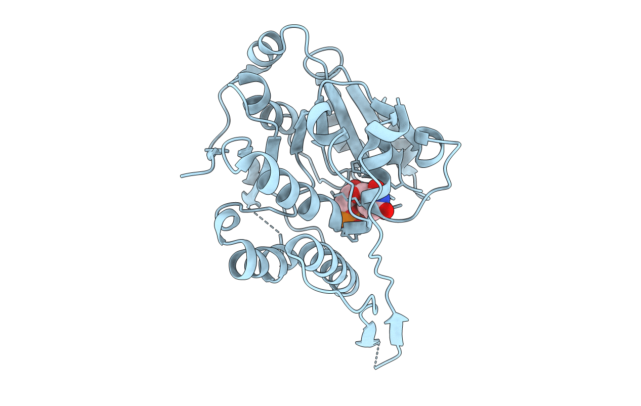
Crystal structure of glucosyl-3-phosphoglycerate synthase from Mycobacterium tuberculosis in complex with uridine-diphosphate (UDP) - GpgS*UDP
Biological Source:
Host Organism:
PDB Version:
Deposition Date:
2016-05-10
Release Date:
2017-05-24
Method Details:
Experimental Method:
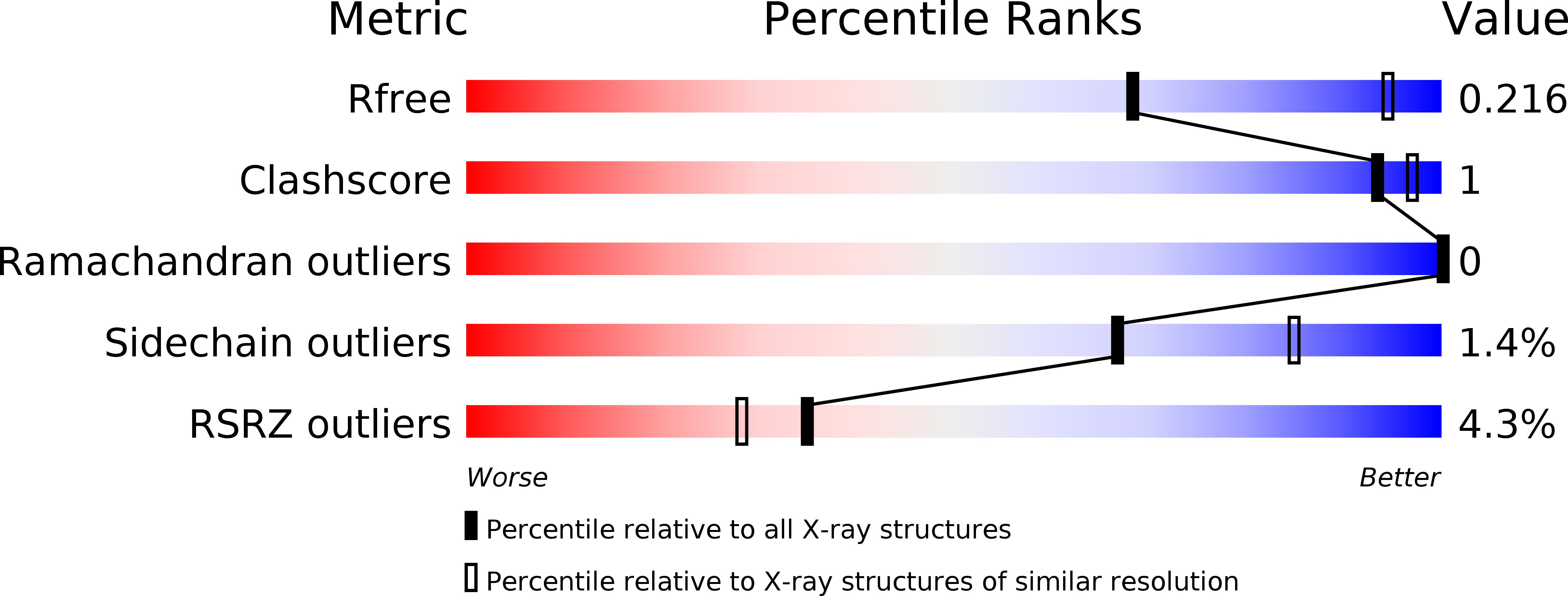
Resolution:
2.59 Å
R-Value Free:
0.21
R-Value Work:
0.19
R-Value Observed:
0.19
Space Group:
I 41
Macromolecular Entities
Polymer Type:polypeptide(L)
Description:Glucosyl-3-phosphoglycerate synthase
Chain IDs:A
Chain Length:328
Number of Molecules:1
Biological Source:Mycobacterium bovis AF2122/97
Ligand Molecules
Primary Citation
Structural Snapshots and Loop Dynamics along the Catalytic Cycle of Glycosyltransferase GpgS.
Structure 25 1034 1044.e3 (2017)
PMID: 28625787 DOI: 10.1016/j.str.2017.05.009

Abstact

Glycosyltransferases (GTs) play a central role in nature. They catalyze the transfer of a sugar moiety to a broad range of acceptor substrates. GTs are highly selective enzymes, allowing the recognition of subtle structural differences in the sequences and stereochemistry of their sugar and acceptor substrates. We report here a series of structural snapshots of the reaction center of the retaining glucosyl-3-phosphoglycerate synthase (GpgS). During this sequence of events, we visualize how the enzyme guides the substrates into the reaction center where the glycosyl transfer reaction takes place, and unveil the mechanism of product release, involving multiple conformational changes not only in the substrates/products but also in the enzyme. The structural data are further complemented by metadynamics free-energy calculations, revealing how the equilibrium of loop conformations is modulated along these itineraries. The information reported here represent an important contribution for the understanding of GT enzymes at the molecular level.

Legend

Protein

Chemical

Disease

Primary Citation of related structures