5JQR image
Deposition Date 2016-05-05
Release Date 2016-12-21
Last Version Date 2024-01-10
Entry Detail
PDB ID:
5JQR
Keywords:
Title:
The Structure of Ascorbate Peroxidase Compound II formed by reaction with m-CPBA
Biological Source:
Source Organism:
Glycine max (Taxon ID: 3847)
Host Organism:
Method Details:
Experimental Method:
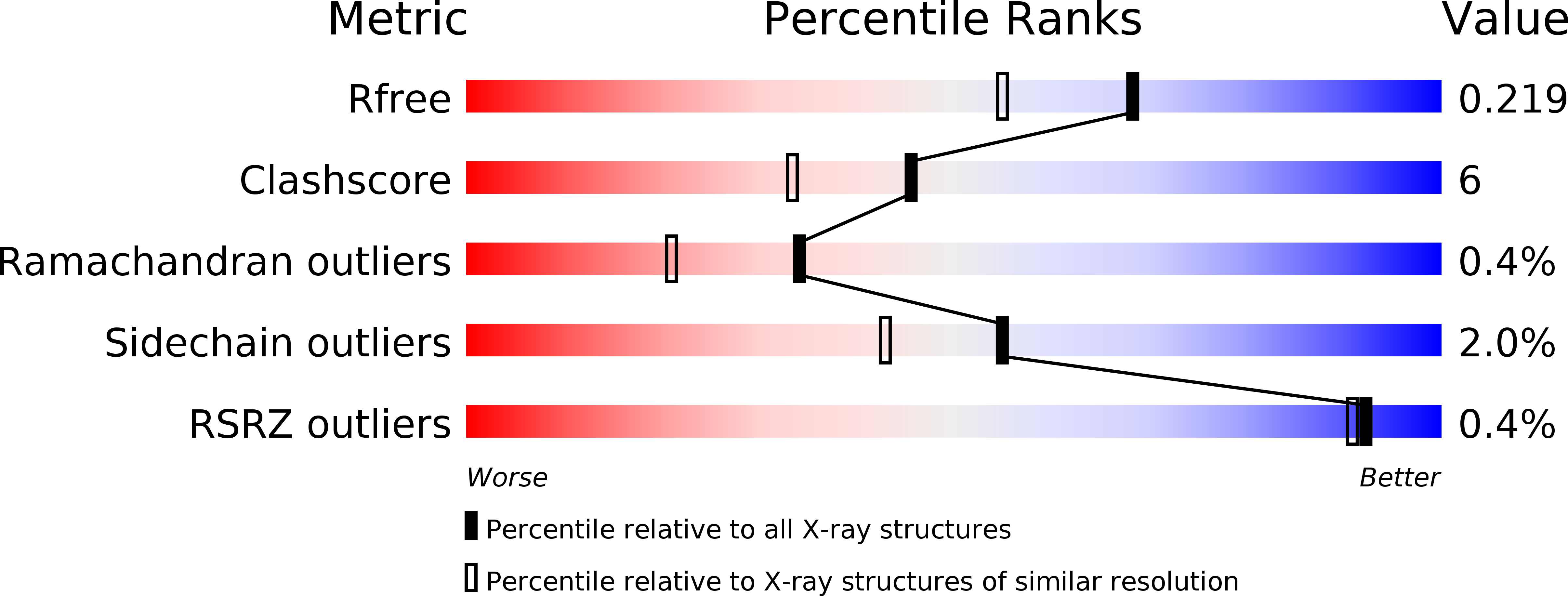
Resolution:
1.81 Å
R-Value Free:
0.21
R-Value Work:
0.15
R-Value Observed:
0.15
Space Group:
P 42 21 2
Macromolecular Entities
Polymer Type:polypeptide(L)
Molecule:Ascorbate peroxidase
Gene (Uniprot):apx1
Chain IDs:A
Chain Length:249
Number of Molecules:1
Biological Source:Glycine max
Primary Citation
Direct visualization of a Fe(IV)-OH intermediate in a heme enzyme.
Nat Commun 7 13445 13445 (2016)
PMID: 27897163 DOI: 10.1038/ncomms13445

Abstact

Catalytic heme enzymes carry out a wide range of oxidations in biology. They have in common a mechanism that requires formation of highly oxidized ferryl intermediates. It is these ferryl intermediates that provide the catalytic engine to drive the biological activity. Unravelling the nature of the ferryl species is of fundamental and widespread importance. The essential question is whether the ferryl is best described as a Fe(IV)=O or a Fe(IV)-OH species, but previous spectroscopic and X-ray crystallographic studies have not been able to unambiguously differentiate between the two species. Here we use a different approach. We report a neutron crystal structure of the ferryl intermediate in Compound II of a heme peroxidase; the structure allows the protonation states of the ferryl heme to be directly observed. This, together with pre-steady state kinetic analyses, electron paramagnetic resonance spectroscopy and single crystal X-ray fluorescence, identifies a Fe(IV)-OH species as the reactive intermediate. The structure establishes a precedent for the formation of Fe(IV)-OH in a peroxidase.

Legend

Protein

Chemical

Disease

Primary Citation of related structures