
Deposition Date
2016-05-04
Release Date
2016-12-21
Last Version Date
2024-05-01
Entry Detail
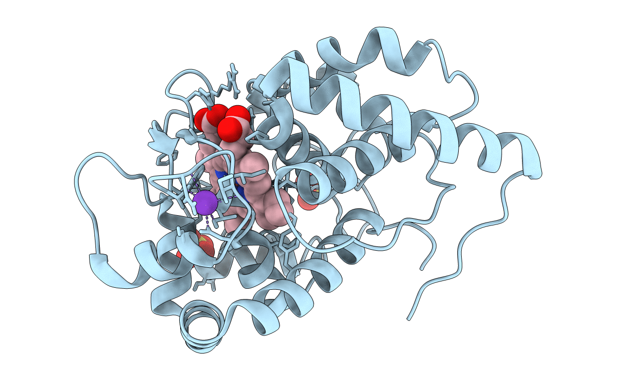
PDB ID:
5JPR
Keywords:
Title:
Neutron Structure of Compound II of Ascorbate Peroxidase
Biological Source:
Source Organism(s):
Glycine max (Taxon ID: 3847)
Expression System(s):
Method Details:
Experimental Method:
R-Value Free:
['0.21
R-Value Work:
['0.15
R-Value Observed:
['0.15
Space Group:
P 42 21 2
