5JM7 image
Deposition Date 2016-04-28
Release Date 2016-06-15
Last Version Date 2024-03-06
Entry Detail
PDB ID:
5JM7
Keywords:
Title:
The structure of aerobactin synthetase IucA from a hypervirulent pathotype of Klebsiella pneumoniae
Biological Source:
Source Organism:
Host Organism:
Method Details:
Experimental Method:
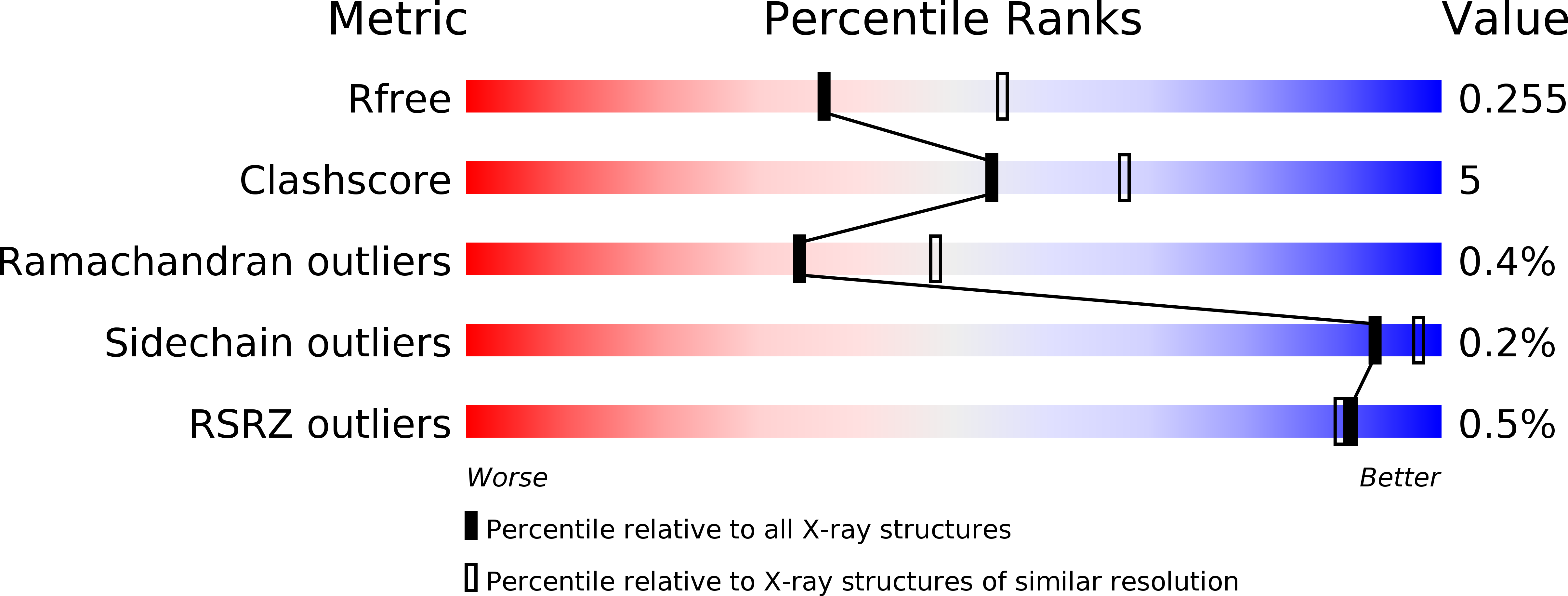
Resolution:
2.40 Å
R-Value Free:
0.25
R-Value Work:
0.21
R-Value Observed:
0.21
Space Group:
P 62 2 2
Macromolecular Entities
Polymer Type:polypeptide(L)
Molecule:Aerobactin synthase IucA
Chain IDs:A
Chain Length:576
Number of Molecules:1
Biological Source:Klebsiella pneumoniae subsp. pneumoniae
Ligand Molecules
Primary Citation
Structural and Functional Characterization of Aerobactin Synthetase IucA from a Hypervirulent Pathotype of Klebsiella pneumoniae.
Biochemistry 55 3559 3570 (2016)
PMID: 27253399 DOI: 10.1021/acs.biochem.6b00409

Abstact

Iron is a vital mineral nutrient required by virtually all life forms to prosper; pathogenic bacteria are no exception. Despite the abundance of iron within the human host, highly regulated iron physiology can result in exceedingly low levels of iron bioavailable to prospective invading bacteria. To combat this scarcity of iron, many pathogenic bacteria have acquired specific and efficient iron acquisition systems, which allow them to thrive in iron-deficient host environments. One of the more prominent bacterial iron acquisition systems involves the synthesis, secretion, and reuptake of small-molecule iron chelators known as siderophores. Aerobactin, a citrate-hydroxamate siderophore originally isolated nearly 50 years ago, is produced by a number of pathogenic Gram-negative bacteria. Aerobactin has recently been demonstrated to play a pivotal role in mediating the enhanced virulence of a particularly invasive pathotype of Klebsiella pneumoniae (hvKP). Toward further understanding of this key virulence factor, we report the structural and functional characterization of aerobactin synthetase IucA from a strain of hvKP. The X-ray crystal structures of unliganded and ATP-bound forms of IucA were solved, forming the foundation of our structural analysis. Small angle X-ray scattering (SAXS) data suggest that, unlike its closest structurally characterized homologues, IucA adopts a tetrameric assembly in solution. Finally, we employed activity assays to investigate the substrate specificity and determine the apparent steady-state kinetic parameters of IucA.

Legend

Protein

Chemical

Disease

Primary Citation of related structures