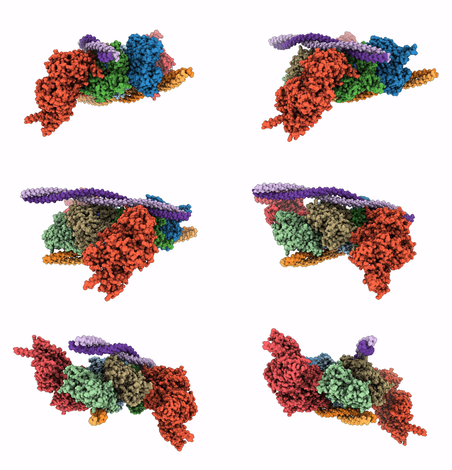
5JLH image
Deposition Date 2016-04-27
Release Date 2016-06-15
Last Version Date 2024-07-10
Entry Detail
PDB ID:
5JLH
Title:
Cryo-EM structure of a human cytoplasmic actomyosin complex at near-atomic resolution
Biological Source:
Source Organism:
Host Organism:
Method Details:
Experimental Method:
Resolution:
3.90 Å
Aggregation State:
FILAMENT
Reconstruction Method:
SINGLE PARTICLE
Macromolecular Entities
Structures with similar UniProt ID
Protein Blast
Polymer Type:polypeptide(L)
Molecule:Actin, cytoplasmic 2
Gene (Uniprot):ACTG1
Chain IDs:A, B, C, D, E
Chain Length:374
Number of Molecules:5
Biological Source:Homo sapiens
Protein Blast
Polymer Type:polypeptide(L)
Molecule:Myosin-14,Alpha-actinin A
Gene (Uniprot):abpA, MYH14
Chain IDs:F, G
Chain Length:1039
Number of Molecules:2
Biological Source:Homo sapiens, Dictyostelium discoideum
Polymer Type:polypeptide(L)
Molecule:Tropomyosin alpha-3 chain
Chain IDs:H, I, J, K
Chain Length:135
Number of Molecules:4
Biological Source:Homo sapiens
Primary Citation
Cryo-EM structure of a human cytoplasmic actomyosin complex at near-atomic resolution.
Nature 534 724 728 (2016)
PMID: 27324845 DOI: 10.1038/nature18295

Abstact

The interaction of myosin with actin filaments is the central feature of muscle contraction and cargo movement along actin filaments of the cytoskeleton. The energy for these movements is generated during a complex mechanochemical reaction cycle. Crystal structures of myosin in different states have provided important structural insights into the myosin motor cycle when myosin is detached from F-actin. The difficulty of obtaining diffracting crystals, however, has prevented structure determination by crystallography of actomyosin complexes. Thus, although structural models exist of F-actin in complex with various myosins, a high-resolution structure of the F-actin–myosin complex is missing. Here, using electron cryomicroscopy, we present the structure of a human rigor actomyosin complex at an average resolution of 3.9 Å. The structure reveals details of the actomyosin interface, which is mainly stabilized by hydrophobic interactions. The negatively charged amino (N) terminus of actin interacts with a conserved basic motif in loop 2 of myosin, promoting cleft closure in myosin. Surprisingly, the overall structure of myosin is similar to rigor-like myosin structures in the absence of F-actin, indicating that F-actin binding induces only minimal conformational changes in myosin. A comparison with pre-powerstroke and intermediate (Pi-release) states of myosin allows us to discuss the general mechanism of myosin binding to F-actin. Our results serve as a strong foundation for the molecular understanding of cytoskeletal diseases, such as autosomal dominant hearing loss and diseases affecting skeletal and cardiac muscles, in particular nemaline myopathy and hypertrophic cardiomyopathy.

Legend

Protein

Chemical

Disease

Primary Citation of related structures