5JK7 image
Deposition Date 2016-04-26
Release Date 2016-10-05
Last Version Date 2024-03-06
Entry Detail
PDB ID:
5JK7
Title:
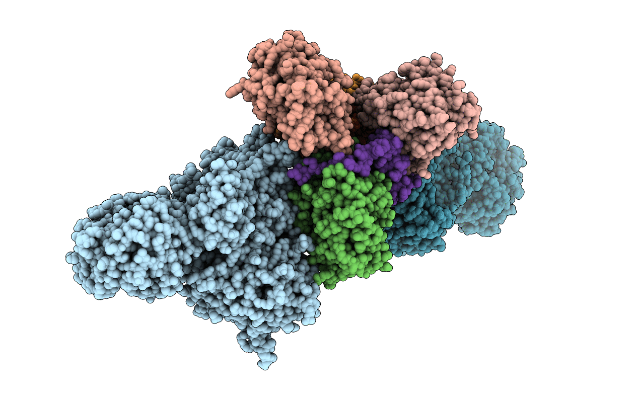
The X-ray structure of the DDB1-DCAF1-Vpr-UNG2 complex
Biological Source:
Method Details:
Experimental Method:
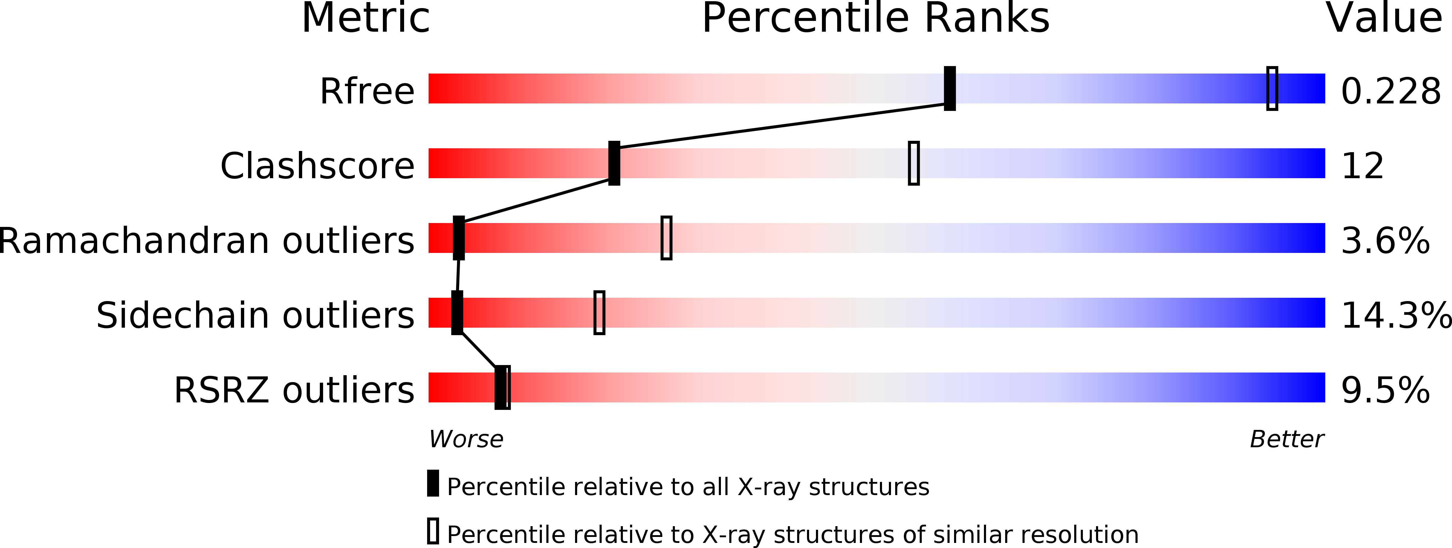
Resolution:
3.49 Å
R-Value Free:
0.20
R-Value Work:
0.17
R-Value Observed:
0.17
Space Group:
P 1
Macromolecular Entities
Structures with similar UniProt ID
Protein Blast
Polymer Type:polypeptide(L)
Molecule:DNA damage-binding protein 1
Gene (Uniprot):DDB1
Chain IDs:A, B
Chain Length:1140
Number of Molecules:2
Biological Source:Homo sapiens
Structures with similar UniProt ID
Protein Blast
Polymer Type:polypeptide(L)
Molecule:Protein VPRBP
Gene (Uniprot):DCAF1
Chain IDs:C, E
Chain Length:361
Number of Molecules:2
Biological Source:Homo sapiens
Structures with similar UniProt ID
Protein Blast
Polymer Type:polypeptide(L)
Molecule:Uracil-DNA glycosylase
Gene (Uniprot):UNG
Chain IDs:D, G
Chain Length:222
Number of Molecules:2
Biological Source:Homo sapiens
Structures with similar UniProt ID
Protein Blast
Polymer Type:polypeptide(L)
Molecule:Protein Vpr
Gene (Uniprot):vpr
Chain IDs:F, H
Chain Length:96
Number of Molecules:2
Biological Source:Human immunodeficiency virus type 1 group M subtype B (isolate NY5)
Ligand Molecules
Primary Citation
The DDB1-DCAF1-Vpr-UNG2 crystal structure reveals how HIV-1 Vpr steers human UNG2 toward destruction.
Nat.Struct.Mol.Biol. 23 933 940 (2016)
PMID: 27571178 DOI: 10.1038/nsmb.3284

Abstact

The HIV-1 accessory protein Vpr is required for efficient viral infection of macrophages and promotion of viral replication in T cells. Vpr's biological activities are closely linked to the interaction with human DCAF1, a cellular substrate receptor of the Cullin4-RING E3 ubiquitin ligase (CRL4) of the host ubiquitin-proteasome-mediated protein degradation pathway. The molecular details of how Vpr usurps the protein degradation pathway have not been delineated. Here we present the crystal structure of the DDB1-DCAF1-HIV-1-Vpr-uracil-DNA glycosylase (UNG2) complex. The structure reveals how Vpr engages with DCAF1, creating a binding interface for UNG2 recruitment in a manner distinct from the recruitment of SAMHD1 by Vpx proteins. Vpr and Vpx use similar N-terminal and helical regions to bind the substrate receptor, whereas different regions target the specific cellular substrates. Furthermore, Vpr uses molecular mimicry of DNA by a variable loop for specific recruitment of the UNG2 substrate.

Legend

Protein

Chemical

Disease

Primary Citation of related structures