5JEV image
Deposition Date 2016-04-19
Release Date 2016-09-14
Last Version Date 2024-05-08
Entry Detail
PDB ID:
5JEV
Keywords:
Title:
del-[Ru(phen)2(dppz]2+ bound to d(TCGGCGCCGA) with Cobalt hexammine
Biological Source:
Source Organism:
Method Details:
Experimental Method:
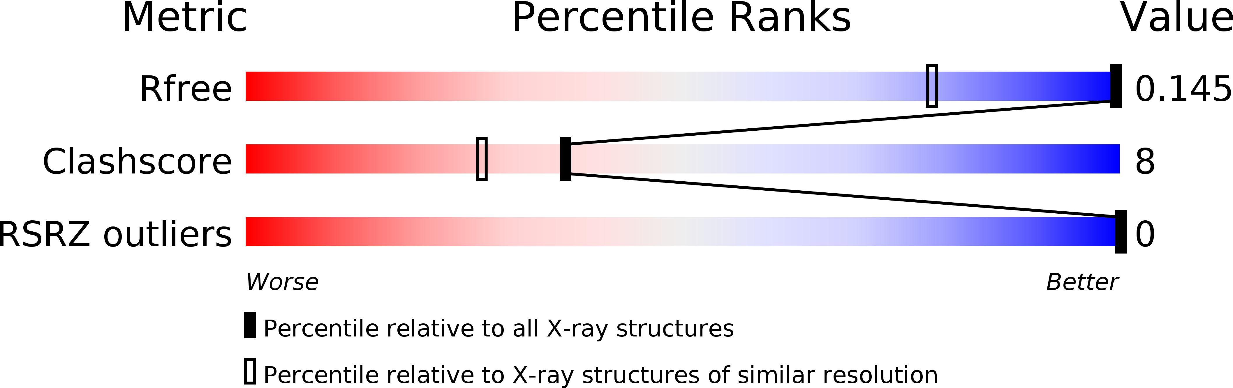
Resolution:
0.99 Å
R-Value Free:
0.14
R-Value Work:
0.12
R-Value Observed:
0.12
Space Group:
P 41 21 2
Macromolecular Entities
Polymer Type:polydeoxyribonucleotide
Molecule:DNA (5'-D(*TP*CP*GP*GP*CP*GP*CP*CP*GP*A)-3')
Chain IDs:A
Chain Length:10
Number of Molecules:1
Biological Source:synthetic construct
Primary Citation
Delta chirality ruthenium 'light-switch' complexes can bind in the minor groove of DNA with five different binding modes.
Nucleic Acids Res. 44 9472 9482 (2016)
PMID: 27599841 DOI: 10.1093/nar/gkw753

Abstact

[Ru(phen)2(dppz)]2+ has been studied since the 1990s due to its 'light-switch' properties. It can be used as a luminescent DNA probe, with emission switched on through DNA binding. The luminescence observed is dependent on the solvent accessibility of the pyrazine nitrogen atoms, and therefore is sensitive to changes in both binding site of the cation and chromophore orientation. The compound is also chiral, and there are distinct differences between the enantiomers in terms of the emission behaviour when bound to a variety of DNA sequences. Whilst a number of binary DNA-complex X-ray crystal structures are available, most include the Λ enantiomer and there is very little structural information about binding of the Δ enantiomer. Here, we present the first X-ray crystal structure of a Δ enantiomer bound to well-matched DNA, in the absence of the other, Λ enantiomer. We show how the binding site observed here can be related to a more general pattern of motifs in the crystallographic literature and propose that the Δ enantiomer can bind with five different binding modes, offering a new hypothesis for the interpretation of solution data.

Legend

Protein

Chemical

Disease

Primary Citation of related structures