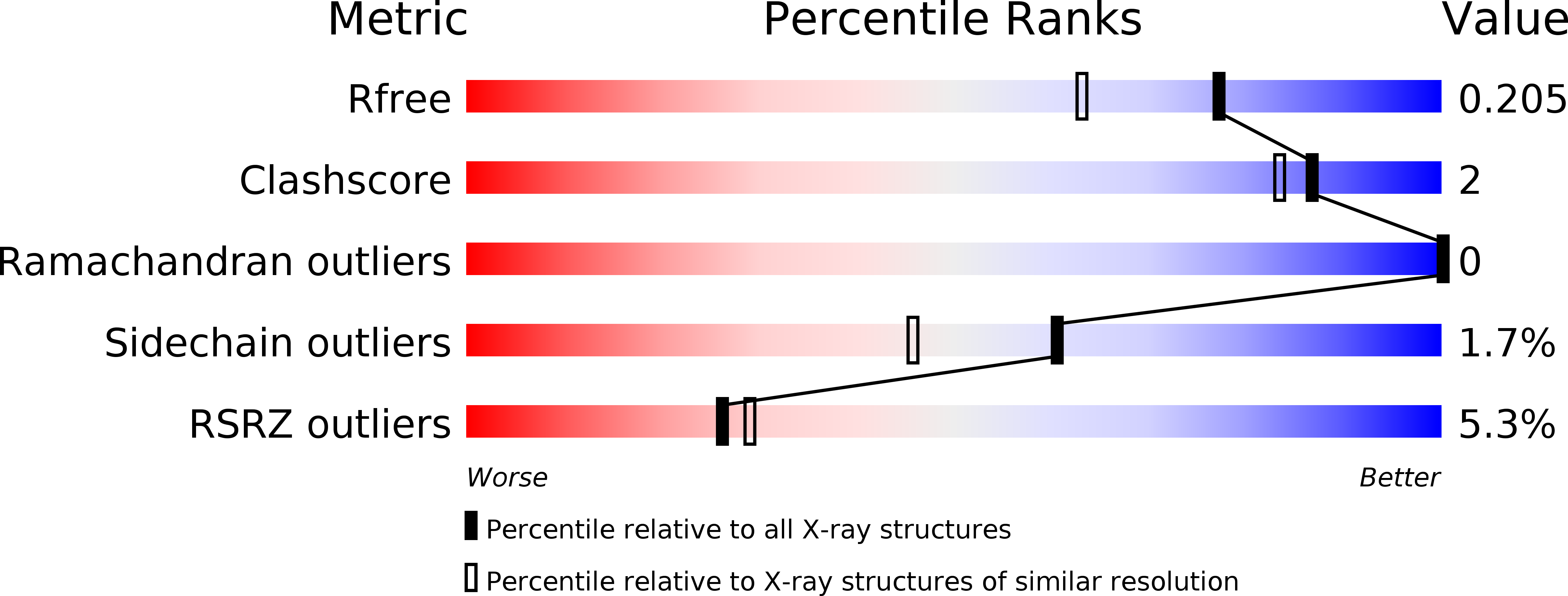
5JDU image
Deposition Date 2016-04-17
Release Date 2016-07-13
Last Version Date 2024-10-30
Entry Detail
PDB ID:
5JDU
Keywords:
Title:
Crystal structure for human thrombin mutant D189A
Biological Source:
Source Organism:
Homo sapiens (Taxon ID: 9606)
Host Organism:
Method Details:
Experimental Method:
Resolution:
1.70 Å
R-Value Free:
0.20
R-Value Work:
0.18
R-Value Observed:
0.18
Space Group:
P 21 21 21
Macromolecular Entities
Structures with similar UniProt ID
Protein Blast
Polymer Type:polypeptide(L)
Molecule:Thrombin light chain
Gene (Uniprot):F2
Chain IDs:A, C
Chain Length:33
Number of Molecules:2
Biological Source:Homo sapiens
Structures with similar UniProt ID
Protein Blast
Polymer Type:polypeptide(L)
Molecule:Thrombin heavy chain
Gene (Uniprot):F2
Mutagens:D189A
Chain IDs:B, D
Chain Length:259
Number of Molecules:2
Biological Source:Homo sapiens
Modified Residue
Compound ID Chain ID Parent Comp ID Details 2D Image
NAG E NAG -D
Primary Citation
Loop Electrostatics Asymmetry Modulates the Preexisting Conformational Equilibrium in Thrombin.
Biochemistry 55 3984 3994 (2016)
PMID: 27347732 DOI: 10.1021/acs.biochem.6b00385

Abstact

Thrombin exists as an ensemble of active (E) and inactive (E*) conformations that differ in their accessibility to the active site. Here we show that redistribution of the E*-E equilibrium can be achieved by perturbing the electrostatic properties of the enzyme. Removal of the negative charge of the catalytic Asp102 or Asp189 in the primary specificity site destabilizes the E form and causes a shift in the 215-217 segment that compromises substrate entrance. Solution studies and existing structures of D102N document stabilization of the E* form. A new high-resolution structure of D189A also reveals the mutant in the collapsed E* form. These findings establish a new paradigm for the control of the E*-E equilibrium in the trypsin fold.

Legend

Protein

Chemical

Disease

Primary Citation of related structures