Deposition Date
2016-04-15
Release Date
2016-11-16
Last Version Date
2024-03-06
Entry Detail
PDB ID:
5JD2
Keywords:
Title:
SFX structure of corestreptavidin-selenobiotin complex
Biological Source:
Source Organism(s):
Streptomyces avidinii (Taxon ID: 1895)
Expression System(s):
Method Details:
Experimental Method:
Resolution:
1.90 Å
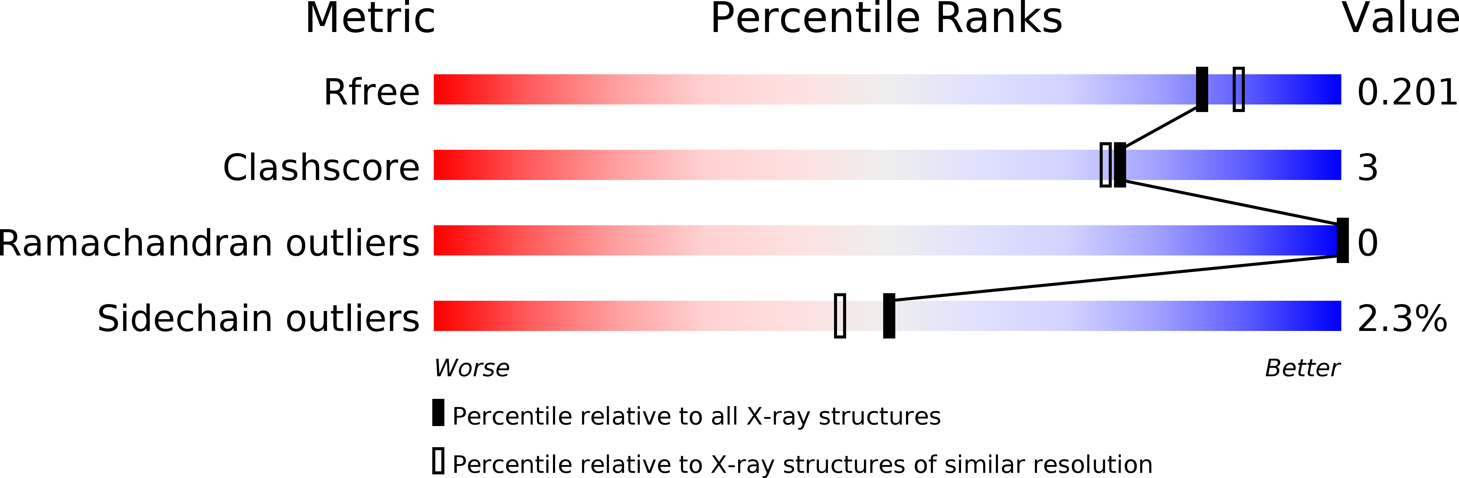
R-Value Free:
0.19
R-Value Work:
0.16
R-Value Observed:
0.16
Space Group:
P 1 21 1