Deposition Date
2016-04-15
Release Date
2016-10-12
Last Version Date
2023-11-08
Entry Detail
PDB ID:
5JCL
Keywords:
Title:
Structure and catalytic mechanism of monodehydroascorbate reductase, MDHAR, from Oryza sativa L. japonica
Biological Source:
Source Organism(s):
Oryza sativa subsp. japonica (Taxon ID: 39947)
Expression System(s):
Method Details:
Experimental Method:
Resolution:
1.80 Å
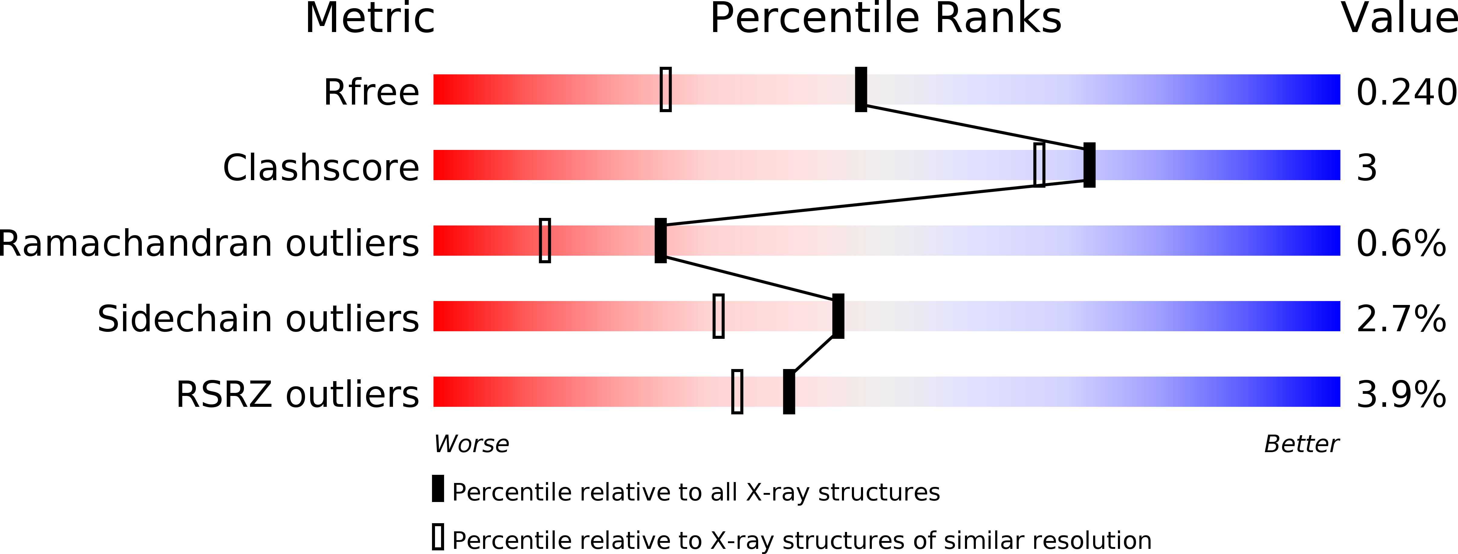
R-Value Free:
0.23
R-Value Work:
0.18
R-Value Observed:
0.18
Space Group:
P 21 21 21